Human Biochemistry Volume 2 - Murray R. 1993
Biochemistry of Intracellular and Intercellular Communications
Sex Gland Hormones
Biosynthesis and Metabolism of Ovarian Hormones
Synthesis
Estrogens are a family of Hormones synthesized in the Ovaries and other Tissues. 17ß-Estradiol is the primary ovarian hormone. In some species, estrone predominates, though it is synthesized extraovarially. During Pregnancy, relatively more estriol is produced, but this hormone is also synthesized outside the ovaries. The primary pathway and subcellular localization of the Enzymes involved in the Initial Stages of estradiol Biosynthesis are identical to those in the Adrenal Glands and Testes. Transformations occurring exclusively in the ovaries are shown in Fig. 50.6.
Estrogens are formed via the aromatization of androgens through a complex process comprising three hydroxylation steps, each requiring O2 and NADPH. Available evidence indicates that the aromatase enzyme complex includes a mixed-function cytochrome P-450 oxidase. When testosterone serves as the substrate for this complex, estradiol is formed; aromatization of androstenedione leads to The production of estrone (a process that typically occurs extraovarially).
Class="center">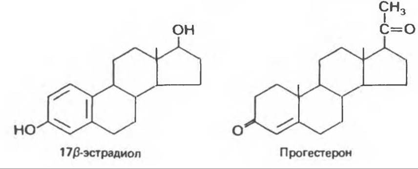
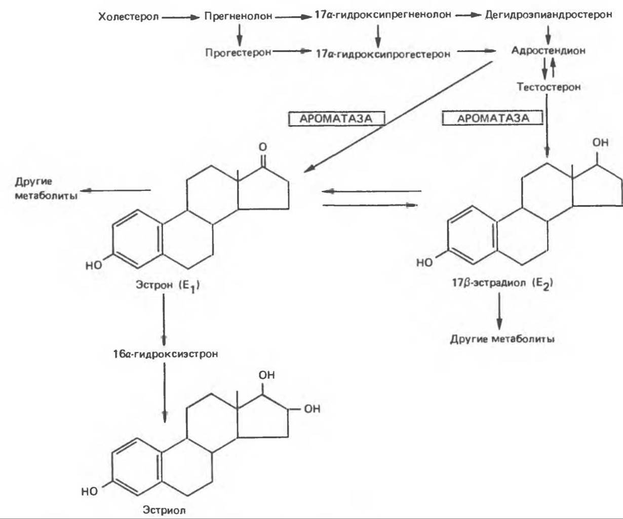
Fig. 50.6. Biosynthesis of estrogens. (Slightly modified and reproduced, with permission, from Ganong W. F. Review of Medical Physiology, 13th ed. Appleton and Lange, 1987.)
It is difficult to pinpoint the exact Cell types responsible for producing Ovarian Hormones. Current understanding suggests that two cell types are involved: 17a-hydroxyprogesterone and androstenedione (the primary androgen produced by the ovaries) are synthesized mainly in the theca Cells, while estradiol is produced in the granulosa cells. Progesterone is produced and secreted by the corpus luteum, which also synthesizes a certain amount of estradiol.
A significant portion of estrogens is formed through peripheral aromatization of androgens. In men, aromatization of testosterone accounts for up to 80% of all estradiol (E2). In women, adrenal androgens serve as an important source of estrogens: up to 50% of E2 produced during pregnancy is formed via the aromatization of DHEA sulfate, while The conversion of androstenedione to estrone serves as the primary source of estrogens in postmenopause. Aromatase activity has been detected in adipose tissue, as well as in the Liver, Skin, and other tissues. Elevated activity of this enzyme appears to play a key role in the "estrogenization" observed in conditions such as liver cirrhosis, hyperthyroidism, Aging, and obesity.
Catechol estrogens are the major estrogen metabolites in all mammals. They are formed by the hydroxylation of the aromatic ring at C-2. Catechol estrogens possess weak estrogenic activity, but they are highly active in the Central Nervous system, where they have also been detected.
Secretion and Transport
The secretion rates of ovarian-derived Steroids fluctuate sharply across the different Phases of the menstrual (or estrous) cycle and depend directly on their rate of production in the ovaries. These hormones are not stored; they are secreted as they are synthesized.
Estrogens and progestins, like Other Steroids, bind to plasma transport Proteins to varying degrees. Estrogens bind to SHBG, whereas progestins bind to CBG. The affinity of SHBG for estradiol is five times lower than for testosterone and DHT; progesterone and cortisol exhibit very low affinity for this protein (Table 50.2). In contrast, progesterone and cortisol bind with nearly equal affinity to CBG, which in turn exhibits low affinity for estradiol and even lower affinity for testosterone, DHT, and estrone.
These plasma transport proteins apparently play no role in the MECHANISM OF ACTION of these hormones at THE CELLULAR LEVEL. Similar to other steroids, likely only the free hormone fraction possesses biological activity. Binding proteins provide a specific hormone reserve in the Blood and, possessing relatively high binding capacity, act as buffers against abrupt fluctuations in plasma hormone levels. The metabolic clearance rates of these hormones are inversely related to their affinity for SHBG. Consequently, the clearance of estrone exceeds that of estradiol, which in turn exceeds that of testosterone or DHT. It should be noted that the conjugated derivatives of these hormones (see below) do not bind to either SHBG or CBG. SHBG fulfills yet another function: due to its differing affinity for estradiol and testosterone (or DHT), it can influence the proportion of these Sex Hormones that interact with target tissues. Factors regulating SHBG production are discussed above.
METABOLISM and Excretion
A. Estrogens. In the liver, estradiol and estrone are converted into estriol via the reactions shown in Fig. 50.6. Estradiol, estrone, and estriol serve as substrates for hepatic enzymes that attach a glucuronide or sulfate group. The activity of these conjugating enzymes varies among species. In rodents, the activity of metabolic enzyme systems (particularly hydroxylating ones) is so high that estrogens are almost completely degraded in The Liver and are virtually inactive when administered orally. In primates, enzymes of this group are less active and, consequently, oral administration of estrogens is more effective in them. Conjugated steroids are Water-soluble and unable to bind to transport proteins. Therefore, they are readily excreted via Bile, feces, and, to a lesser extent, urine.

Fig. 50.7. Biosynthesis of progesterone and its principal metabolic pathway. Other metabolites have also been identified. (Slightly modified and reproduced, with permission, from Ganong W. F. Review of Medical Physiology, 13th ed. Appleton and Lange, 1987.)
B. Progestins. Progesterone is ineffective when administered orally because it is rapidly metabolized in the liver to yield a series of compounds. Sodium pregnanediol-20-glucuronide is the primary progestin metabolite detected in human urine (Fig. 50.7). Certain synthetic steroids, such as 17a-hydroxyprogesterone and 17a-alkyl-substituted 19-nortestosterone compounds, exhibit progestational activity while remaining immune to hepatic transformations. Consequently, they are widely used as oral contraceptives.
Last update: 06/08/2026
Editorial and Educational Adaptation: This material has been compiled based on the primary/original source text. The project team performed an editorial review, corrected technical inaccuracies, structured sections, and adapted the content for an educational format.
What was processed:
- elimination of formatting defects (OCR errors, structural breaks, corrupted characters);
- editorial organization of content;
- standardization of terminology in accordance with academic sources;
- verification of factual statements against the original source text.
All mentions of the author, publication year, and origin of the primary text have been preserved in accordance with the source.