LEHNINGER PRINCIPLES OF BIOCHEMISTRY - VOL. 2. BIOENERGETICS AND METABOLISM - 2014
PART II. BIOENERGETICS AND METABOLISM
19. OXIDATIVE PHOSPHORYLATION AND PHOTOPHOSPHORYLATION
19.7. Light Absorption
Visible light is electromagnetic radiation with wavelengths ranging from 400 to 700 nm, which constitutes a small fraction of the solar radiation spectrum extending from violet to red (Fig. 19-46). The energy of photons (quanta of light energy) is inversely proportional to the wavelength of light. Photons in the violet region of the visible spectrum (short-wavelength, higher-frequency) carry significantly more energy than those in the red region. The energy (E) of a single photon in the visible spectrum can be calculated using Planck's equation:
Е = hv = hc/λ
where h is Planck's constant, h = 6.625 • 10-34 J • s, v is the wavenumber or frequency in s-1, c is the speed of light, c = 3.00 • 108 m/s, and λ is the wavelength (in meters). The energy of a visible light photon varies depending on its wavelength (frequency): about -150 kJ/einstein in the red region and about -300 kJ/einstein in the violet region.
Class="center">Fig. 19-46. Electromagnetic radiation. The electromagnetic radiation spectrum and photon energy in the visible region. 1 Einstein = 6 • 1023 photons.

The ability of a chemical compound to absorb light depends on the distribution of electrons around the nuclei in its molecule. When a molecule (a chromophore) absorbs a single photon, one of its electrons is promoted to a higher energy level. This occurs in an "all-or-none" fashion: to elevate an electron to a higher energy level, the photon must possess a specific amount of energy (a quantum, from the Latin quantum, meaning how much) that precisely matches the energy difference of the electronic transition. A molecule that has absorbed a photon enters a high-energy, excited state, which is typically unstable. If the light source is removed, electrons in high-energy orbitals usually rapidly drop back to their low-energy levels, returning the molecule to its initial stable state, known as the ground state, while releasing the excitation energy (as light or heat). Fluorescence is the light emitted by an excited molecule as it returns to the ground state; the wavelength of the emitted fluorescent light is always longer than that of the absorbed light (see Box 12–3, Vol. 1). Another pathway by which an excited molecule returns to its ground state—crucial for Photosynthesis—involves the direct transfer of excitation energy from the excited molecule to a neighboring molecule in the ground state. This energy transfer process is called exciton transfer, and the quantum of energy transferred from one molecule to another is called an exciton, by analogy with the photon, the quantum of light energy.
Example 19-2 Photon Energy
Vascular plants utilize light with a wavelength of about 700 nm in photosynthesis. Calculate the energy of a "mole" of photons (an einstein of photons) of this wavelength, and compare this value with the energy required to synthesize 1 mol of ATP.
Solution. The energy of a single photon is determined by Planck's equation. At a wavelength of 700 • 10-9 m, the energy of a single photon is
Е = hc /λ = [(6.626 • 10-34 J • s)(3.00 • 108 m/s)]/(7.00 • 10-7 m) = 2.84 •10-19 J
1 einstein = 1 mole of photons, which corresponds to the number of photons equal to Avogadro's number (6.022 • 1023). From this, we can calculate the energy of one einstein of photons at 700 nm:
(2.84 • 10-19 J/photon) (6.022 • 1023 photons/einstein) = 17.1 • 104 J/einstein = 171 kJ/einstein
Thus, 1 mole of red light photons possesses energy that is approximately 5 times greater than the energy required to form 1 mole of ATP from ADP and Pi (30.5 kJ/mol).
Chlorophylls absorb light energy in photosynthesis
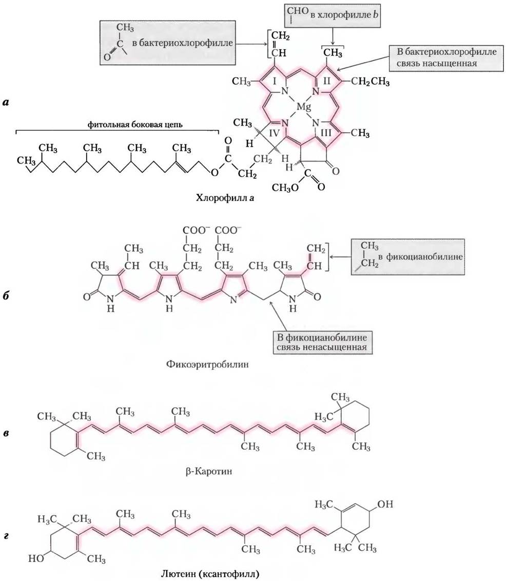
Let us now examine the light-absorbing pigments of the thylakoid membranes. The primary among them are the green pigments, chlorophylls —Mg2+-containing molecular complexes with a polycyclic planar Structure similar to the protoporphyrin of Hemoglobin, except that the coordinating ion is Mg2+ rather than Fe2+ (see Fig. 5–1, Vol. 1). Four nitrogen atoms pointing toward the center of the ring structure are coordinatively bonded to the Mg2+ ion (Fig. 19-47). All chlorophylls feature a long isoprenoid side chain—the residue of the alcohol phytol, esterified to a carboxyl group of a substituent on ring IV. Unlike Hemes, chlorophylls possess a fifth (non-pyrrole) ring.
The five-membered heterocycles surrounding the Mg2+ ion form a conjugated system of alternating single and double bonds, which gives rise to intense characteristic absorption bands in the visible spectrum that are essential for photosynthesis (Fig. 19-48). Chlorophylls have unusually high extinction coefficients (see Box 3–1, Vol. 1), meaning they absorb visible light very efficiently during photosynthesis.
METABOLISM/14.html">Chloroplasts always contain Two Types of chlorophyll—a and b (Fig. 19-47a). Although both are green, their absorption spectra differ slightly (Fig. 19-48). In most higher plants, The amount of chlorophyll a is approximately twice that of chlorophyll b. The chlorophylls of Algae and photosynthetic Bacteria differ slightly from those of higher plants.
Chlorophyll molecules are invariably associated with specific Proteins, forming Light-Harvesting Complexes in which the pigment molecules are precisely oriented relative to one another and to other Protein Complexes within the thylakoid membrane. One such light-harvesting complex (type II; Fig. 19-49) contains seven molecules of chlorophyll a, five molecules of chlorophyll b, and two molecules of the accessory pigment lutein (described below).
In cyanobacteria and red algae, light energy receptors are represented by a different class of pigments—the red and blue phycobilins (phycoerythrobilins and phycocyanobilins, respectively; Fig. 19-47b). Phycobilins are open-chain tetrapyrroles with an extensive system of conjugated double bonds. Unlike chlorophyll, phycobilins contain no Mg2+ and lack a cyclic tetrapyrrole structure. Complexes of phycobilins with specific proteins are called phycobiliproteins. They are organized into large supramolecular assemblies known as phycobilisomes (Fig. 19-50), which serve as the primary light-harvesting systems in these microorganisms.
Accessory pigments broaden the wavelength range of absorbed light
In addition to chlorophylls, Thylakoid membranes contain minor or accessory light-harvesting pigments. These accessory pigments include various colored carotenoids ranging from yellow to red and purple. Among the most important are the red isoprenoid pigment β-carotene and the yellow carotenoid lutein (Fig. 19-47c and d). Carotenoids absorb light in wavelength ranges different from those of chlorophylls and thus function as complementary light receptors that augment chlorophyll absorption (Fig. 19-48).
Fig. 19-47. Major and accessory photopigments: (a) chlorophylls a and b and bacteriochlorophyll are the primary light-absorbing pigments; (b) phycoerythrobilin and phycocyanobilin (the phycobilin group) serve as antenna, or light-harvesting, pigments in cyanobacteria and red algae; (c) β-carotene and other carotenoids, as well as lutein (a xanthophyll), act as accessory pigments in green leaves. All these pigments contain a system of conjugated bonds that enables them to absorb light in the visible region (highlighted in pink).
Experimental data on photosynthetic efficiency as a function of wavelength are presented as a photochemical action spectrum (Fig. 19-51). This spectrum can also be used to identify the primary pigments responsible for the biological activity of light of a specific wavelength. By being able to absorb light energy within a defined wavelength range, Photosynthetic organisms are able to occupy unique ecological niches. For example, the phycobilin pigments of red algae and cyanobacteria absorb light in the 520–630 nm range (Fig. 19-48), allowing them to thrive surrounded by organisms whose pigments—and Water itself—absorb light at shorter or longer wavelengths.
Fig. 19-48. Absorption spectra of photopigments in the visible region. Plants appear green because their pigments absorb visible light in the red and blue regions while reflecting or transmitting green light. Comparing the solar radiation spectrum reaching the Earth's surface with the absorption spectra of plant pigments shows that chlorophylls a and b, along with accessory pigments, enable plants to capture the bulk of the solar energy arriving on Earth. The ratio of chlorophylls to various carotenoids varies markedly among plant species, determining the characteristic green color of photosynthetic Cells. This color ranges from the blue-green of spruce needles or the bright green of maple leaves to the red, brown, or even purple hues found in various multicellular algae and the leaves of certain ornamental plants.

Fig. 19-49. STRUCTURE OF THE light-harvesting complex type II (PDB ID 2BHW). The functional light-harvesting complex is a trimer containing 36 chlorophyll molecules and 6 lutein molecules. The monomer shown here (viewed in the plane of the membrane) consists of three transmembrane α-helical segments, seven molecules of chlorophyll a (light green), five molecules of chlorophyll b (dark green), and two molecules of the accessory pigment lutein (yellow), which form the internal framework.

Fig. 19-50. Structure of a phycobilisome. The phycobilisome of cyanobacteria and red algae consists of phycobilin complexes associated with specialized proteins: phycoerythrin (PE), phycocyanin (PC), and allophycocyanin (APC). Photon energy absorbed by phycoerythrin and phycocyanin is rapidly transferred as an exciton via the phycocyanobilin-allophycocyanin-chlorophyll a protein complex to the reaction center. This process is discussed in more detail in the text.

Fig. 19-51. Two Methods FOR STUDYING the photochemical action spectrum: (a) Results of the classical 1882 experiment by T. W. Engelmann to determine the wavelengths most effective for photosynthesis. Filaments of a photosynthetic marine alga were placed on a Microscope slide and illuminated with light dispersed through a prism, so that one section of the filaments received blue light, another yellow, and a third red. Bacteria known to migrate toward regions of highest oxygen concentration were added to the slide. The effectiveness of visible light at a given wavelength was assessed by the number of bacteria accumulating in each region—violet and red light produced the best results. (b) Results of a similar experiment, differing in that the amount of oxygen evolved during photosynthesis was measured using an oxygen electrode. The photochemical action spectrum shows the dependence of relative photosynthetic efficiency on wavelength. Comparing the absorption spectra of the pigments with the photochemical action spectrum of photosynthetic cells allows for an evaluation of the light-absorbing Properties of the pigments (see Fig. 19-48).

Chlorophylls transfer light energy to reaction centers via exciton transfer to neighboring molecules
The light-absorbing pigments of the thylakoid membranes are organized into functional complexes called Photosystems. For example, the photosystem in spinach chloroplasts contains about 200 chlorophyll molecules and roughly 50 carotenoid molecules. While all pigment molecules absorb photons, only specific chlorophyll molecules can convert light into chemical energy. These specialized chlorophylls are bound to specific proteins to form a photochemical reaction center. All other pigment molecules are termed light-harvesting or antenna pigments. They absorb light and rapidly transfer its energy to the photochemical reaction centers, where the photochemical reaction takes place (Fig. 19-52).
Fig. 19-52. Organization of a photosystem in the thylakoid membrane. Thylakoid photosystems consist of several hundred antenna chlorophyll molecules and accessory pigments oriented in a specific geometry relative to the photochemical reaction center. The absorption of a photon by an antenna chlorophyll molecule raises that molecule to an excited state. The excitation energy (an exciton) is then rapidly transferred from pigment molecule to pigment molecule toward the reaction center (the exciton pathway is indicated by the black arrow). Thylakoid membranes also house the cytochrome b6f complex and ATP synthase (see Fig. 19-60).

The light-absorbing properties of chlorophyll molecules in photochemical reaction centers differ markedly from those of isolated molecules. When a chlorophyll solution is illuminated in vitro, the absorbed energy is rapidly lost as fluorescence or dissipated as heat. In contrast, chlorophyll in intact leaves transitions to an excited state upon exposure to visible light with very low fluorescence emission (Fig. 19-53, step (1)). Instead, a sequence of events unfolds stepwise within the chloroplasts. An excited antenna chlorophyll molecule transfers its energy directly to a neighboring molecule, raising it to an excited state while returning itself to the ground state (step (2)). The excitation energy (exciton) then rapidly migrates through subsequent pigment molecules (third, fourth, etc.) to the photochemical reaction center, where a single chlorophyll molecule within a special pair of type a chlorophylls becomes excited (step (3)). In this excited chlorophyll molecule, an electron is promoted to a higher energy orbital. This excited electron leaves the reaction center chlorophyll and is transferred to the primary electron acceptor in the Electron Transport Chain, leaving behind a "hole" in the reaction center, conventionally represented in diagrams by a "+" sign (Fig. 19-53) (step (4)). The primary electron acceptor becomes reduced upon accepting the electron, whereas the reaction center becomes oxidized upon losing its electron. The loss of an electron by the reaction center chlorophyll is replenished by an electron from a neighboring electron-donating molecule, which thereby acquires a positive charge (step (5)). Thus, light energy absorbed by chloroplasts drives a series of oxidation-reduction reactions.
Fig. 19-53. Pathway of excitons and electrons in chloroplasts. The conversion of absorbed photon energy into charge Separation within the photochemical complexes of chloroplasts occurs in several stages. Step (1) is repeated among the antenna molecules until the exciton reaches a reaction center chlorophyll and excites it. For details on the subsequent states, see the text. The asterisk denotes the excited state of the molecule.

Summary of Section 19.7 Light absorption
■ Photophosphorylation in the chloroplasts of green plants and cyanobacteria is coupled to Electron transport along a chain of carriers.
■ During the light reactions of photosynthesis in green plants, absorbed light energy excites chlorophyll and accessory pigment molecules, which subsequently transfer this excitation energy to reaction centers in the thylakoid membranes. Within the reaction centers, photoexcitation generates electrical charges, producing a strong electron donor (reductant) and a strong electron acceptor (oxidant).
Last update: 06/08/2026
Editorial and Educational Adaptation: This material has been compiled based on the primary/original source text. The project team performed an editorial review, corrected technical inaccuracies, structured sections, and adapted the content for an educational format.
What was processed:
- elimination of formatting defects (OCR errors, structural breaks, corrupted characters);
- editorial organization of content;
- standardization of terminology in accordance with academic sources;
- verification of factual statements against the original source text.
All mentions of the author, publication year, and origin of the primary text have been preserved in accordance with the source.