ECOLOGICAL BIOCHEMISTRY - Textbook - V. M. Isaenko 2005
Chapter 2. ECOLOGICAL AND BIOCHEMICAL INTERACTIONS OF PROKARYOTES, MICROSCOPIC FUNGI, AND ALGAE WITH HIGHER PLANTS AND ANIMALS
2.4. Mechanisms of microbial action on plants
The relationships between microorganisms and higher plants can take A wide variety of forms. One such form is mutually beneficial for the higher plant. Typical Examples of this are the well-studied mycosis and rhizosphere.
Mycosis (from Greek *mykes* — fungus) is a mutually beneficial coexistence (Symbiosis) between fungal mycelium and the roots of higher plants, formed mainly by Basidiomycetes (agaric and bolete mushrooms), as well as certain zygomycetes and ascomycetes. Mycorrhiza is widespread among various groups of higher plants, primarily woody species. It also occurs in perennial grasses and, more rarely, in annuals.
In ectotrophic mycorrhiza, the fungus (mycelium) envelops the ROOT and remains on its surface (for example, the symbiosis of basidiomycetous Fungi with forest trees). Endotrophic mycorrhiza is characterized by the penetration of the fungus into the root Tissues (microscopic fungi with plants of the orchid and heather families). A transitional or ectendotrophic mycorrhiza also occurs. Mycorrhiza can take the form of mutualistic symbiosis, where both Higher Plants and fungi benefit, or exist as a form of restricted parasitism.
The Physiological Role of mycorrhiza is that microscopic fungi provide higher plants with mineral Nutrition and Organic compounds, and help regulate their Water regime. Fungi produce growth activator- and Vitamin-like substances for higher plants, while consuming CARBOHYDRATES from the plant roots.
Fungi that form mycorrhizae in trees only spread under specific biocenotic conditions. Therefore, in forestry and horticulture, saplings are inoculated with microflora. To do this, soil from where the plant previously grew is added when planting trees.
A layer of soil (2–3 cm) directly adjacent to plant roots has an elevated concentration of microorganisms. This soil layer is called the rhizosphere (from Greek *rhiza* — root and *sphaira* — sphere/layer). The composition of rhizosphere microorganisms depends on the soil type, as well as the species and age of the plants. It is enriched with root exudates and decaying root hairs, which provide nutrition for the microorganisms.
Rhizosphere microorganisms are capable of converting certain soil substances into forms readily accessible to plants, synthesizing BIOLOGICALLY ACTIVE SUBSTANCES, enhancing the absorptive capacity of The Root System, stimulating and activating it, and participating in denitrification, among other processes.
Another type of mutually beneficial plant-fungus association can be seen in perennial ryegrass (*Lolium perenne*). The resistance of this plant to the harmful factors of *Listonotus bonariensis* is due to the presence of the so-called Lolium endophyte toxin in the plant Organism, which is produced by the fungus *Acremonium coenophialum*. In plants of the fescue *Festuca orientalis*, which lives in symbiosis with an identical fungal endophyte, pyrrolizidine Alkaloids N3-acetylloline and N-formylloline have been found.
Zoospores of pathogenic fungi are attracted to plant roots with the help of attractants (see Chapter 3.4). The Chemical Structure of A number of such attractants has been established. For instance, in *Aphanomyces euteiches*, it is 5,4'-dihydroxy-7-methoxyisoflavone (prunetin) (Fig. 2.2).
THE SPECTRUM OF prunetin's biological action is very broad: estrogenic and antioxidant activity, stimulation of indole-3-acetic acid oxidase activity, and more.
Class="center">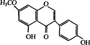
Fig. 2.2. Structure of prunetin from the roots of pea (*Pisum sativum*), which acts as an attractant for *Aphanomyces* zoospores
Pathophytotoxins. Parasitic microorganisms possess a wide range of damaging factors affecting higher plants. One of these is the production and secretion of pathophytotoxins. A significant number of bacterial and fungal pathophytotoxins have been characterized to date.
Low-molecular-weight pathophytotoxins generally affect Plant Growth and cause diseases leading to wilting of leaves and shoots (known as wilt), whereas high-molecular-weight ones cause plant necrosis, tissue maceration (Cell Separation), and so on. Both low- and high-molecular-weight pathophytotoxins can be produced by the same organisms. For example, Dutch elm disease is caused by the fungus *Ceratocystis ulmi*, which is vectored by insects, specifically the bark beetle *Scolytus multistriatus*. The fungus secretes several pathophytotoxins that cause necrotic lesions on leaves and shoots, as well as their wilting. These toxins represent a mixture of Glycoproteins and low-molecular-weight phenolic metabolites (Fig. 2.3).
Fungi of various *Fusarium* species (particularly *Fusarium oxysporum*) produce mycomarasic acid and fusaric acid (Fig. 2.3), which cause wilt in tomatoes. The first of these pathophytotoxins exhibits metal-chelating properties; its translocation and functioning are associated with The formation of a water-soluble complex with iron. The production of fusaric acid depends on the availability of zinc in the environment—its synthesis is suppressed in the case of a zinc deficiency.
Fusaric acid, besides tomatoes, has also been detected in other plants (cotton, flax, etc.) infected with various *Fusarium* species.
Picolinic acid (Fig. 2.3), much like fusaric acid, acts as a metal chelator, binding Iron and copper ions, which leads to The Development of diseases such as rice blast. This acid is detoxified by resistant rice varieties through the formation of complex methyl esters.
In addition to toxic phenols that cause Dutch elm disease, pyridine derivatives that lead to tomato wilt and rice blast, There is a whole range of other pathophytotoxins that induce plant diseases. Among them are cyclic Peptides (e.g., tentoxin from
*Alternaria tenuis*), certain naphthoquinones, and most terpenoids (e.g., helminthosporoside from *Helminthosporium sativum*) (Fig. 2.3).

Fig. 2.3. Structure of low-molecular-weight pathophytotoxins produced by fungi:
1 — pathophytotoxins of *Ceratocystis ulmi* R=CH2COCH3; CHOHCOCH3 or COCOCH3 (Dutch elm disease); 2 — picolinic acid, R = H (*Pyricularia oryzae*, rice blast), fusaric acid, R = C4H9 — butyl (*Fusarium oxysporum*, tomato wilt); 3 — tentoxin (*Alternaria tenuis*, cotton chlorosis); 4 — helminthosporoside (*Helminthosporium sativum*, sugarcane spot blotch): = Gal—galactose
Microorganisms are capable of infecting plants even without producing phytotoxins. As already known, they sometimes disrupt plant cell METABOLISM, leading, for example, to the excessive accumulation of a primary metabolite. For instance, the fungus Rhizopus (family Mucoraceae) can induce the accumulation of fumaric acid in the plant mesocarp. It is then transported to the leaves and shoots, where it is oxidized to form toxic epoxysuccinic acid. The next step involves The conversion of this toxin into tartaric acid, and subsequently into oxaloacetic acid, which enters the Krebs cycle (Tricarboxylic Acid Cycle) (Fig. 2.4).
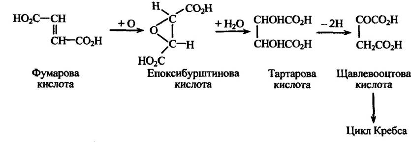
Fig. 2.4. Metabolism of fumaric acid, during which toxic epoxysuccinic acid is formed
Thus, in this case, an excess of a metabolite (fumaric acid) leads to the production of a phytotoxin—epoxysuccinic acid—which is capable of damaging plant Cells.
High-molecular-weight phytotoxins are generally highly toxic even at low concentrations. One such substance is victorin, produced by Helminthosporium victoriae, which affects oat plants. Other similar high-molecular-weight phytotoxins have been isolated from the tissues of pea, corn, and sorghum.
Symptoms that can be directly associated with the action of phytotoxins also include chlorosis and necrosis. Chlorosis (the destruction of Chloroplasts) leads to the loss of the green color in leaves. The same effect is caused by the accumulation of ammonia in the leaves. For example, the toxin produced by the tobacco wildfire pathogen, the bacterium Pseudomonas tabaci, is a small peptide that inhibits glutamate synthetase. As a result, the ammonia generated during nitrate reduction fails to react with glutamic acid and accumulates.
Plant growth abnormalities are in most cases caused by an increased synthesis of growth Hormones, which alter metabolism and stimulate rapid longitudinal growth in herbaceous plants. Stems become elongated and bend under their own weight, leading to lodging. They weaken and become susceptible to saprophytic fungi. An example of a pathogen producing this effect in rice is the fungus Gibberella fujikuroi. Gibberellins, named after this fungus, are also produced by other fungal species, notably from the genus Taphrina.
The synthesis of growth hormones has been detected during the infection of peas by Corynebacterium fascians, where cytokinin production leads to fasciations. Indole-3-acetic acid (auxin), produced by Agrobacterium tumefaciens, induces crown gall tumors in plants.
A common symptom of the disease is necrosis—the appearance of dark, dry, and brittle patches in leaf tissues. One of the possible causes of necrosis is the inhibition of primary metabolism. For instance, when apple trees are infected by the bacterium Erwinia amylovora, ammonia is released, causing SHOOT necrosis.
Among the fungal Enzymes that degrade Plant Cell Walls, the following can be distinguished: 1) pectinases (pectin methylases, pectinesterases, polygalacturonases, etc.), which disrupt the bonds between Cellulose microfibrils and The Cell wall; 2) enzymes that degrade cellulose fibrils; 3) enzymes that hydroxylate other Polysaccharides (Xylan, galactomannan, arabinogalactan, etc.); and 4) enzymes that degrade Lignin.
A number of fungi produce Phosphatases, dehydrogenases, peroxidases, and Other Enzymes that modify and break down the substances plants use to defend themselves against fungal attacks.
Fungal enzymes play a biospheric role as decomposers that break down organic matter, thereby ensuring The breakdown of polymers within ecosystems.
In addition to producing enzymes, microorganisms also synthesize Enzyme Inhibitors that regulate enzyme activity and protect fungal cells from their own enzymes as well as from those of other fungi and higher plants.
One of the mechanisms of plant infection that causes vascular wilt is the clogging of water- and nutrient-conducting vessels. This occurs because certain fungi produce polysaccharide resins that block the xylem, thereby restricting the plant's water uptake. For example, alongside phytotoxins, the fungus Fusarium oxysporum produces vasinfuscarin, which plugs the plant's Vascular System. Bacteria are also capable of blocking plant vessels with their mucous masses.
Sometimes phytotoxins can directly affect plant water relations. For instance, they are capable of interfering with the hormonal control of leaf stomatal function.
Last update: 06/08/2026
Editorial and Educational Adaptation: This material has been compiled based on the primary/original source text. The project team performed an editorial review, corrected technical inaccuracies, structured sections, and adapted the content for an educational format.
What was processed:
- elimination of formatting defects (OCR errors, structural breaks, corrupted characters);
- editorial organization of content;
- standardization of terminology in accordance with academic sources;
- verification of factual statements against the original source text.
All mentions of the author, publication year, and origin of the primary text have been preserved in accordance with the source.