Biochemistry and Molecular Biology - Belyasova N.A. 2002
Metabolism. Energy-Yielding Processes
Respiration
Mechanism of Oxidative Phosphorylation
The entire carrier system within the Respiratory Chain of The inner mitochondrial membrane comprises approximately 70 different Polypeptides organized into four enzyme complexes: NADH—CoQ reductase (complex I), CoQH2—cytochrome c reductase (complex III), cytochrome c oxidase (complex IV), and succinate—CoQ reductase (complex II). The respiratory chain is closely associated with the ATP synthase complex, where ADP phosphorylation takes place. To explain how Electron transport along the respiratory chain components is coupled with ATP synthesis, several hypotheses have been proposed:
1) the chemical coupling hypothesis (E. Slater, 1953): electron transport triggers a series of Chemical Reactions resulting in The formation of a high-energy intermediate. The Cleavage of this product is accompanied by the release of sufficient energy to synthesize an ATP molecule;
2) the conformational coupling hypothesis (P. Boyer, 1964): electron transport induces Conformational Changes in membrane protein components, shifting them into a high-energy state. These changes are transmitted to ATP synthase, which becomes activated and catalyzes ATP synthesis;
3) the chemiosmotic hypothesis (proposed by Peter Mitchell in 1961): Electron transport is accompanied by the extrusion of protons into the mitochondrial intermembrane space. This establishes an electrochemical gradient across the inner membrane, which drives the operation of ATP synthase. In other words, ATP synthesis is powered by the osmotic energy of the proton gradient.
Among all the hypotheses considered, Mitchell's hypothesis is the most robust and supported by extensive experimental data. In particular, it explains the necessity of membrane integrity for ATP synthesis. However, its tenets still hold the status of a theory and are far from perfect, although accepted by the majority of researchers and discussed below.
Each complex of the respiratory chain includes several redox centers positioned within the membrane in a specific manner to form several 'loops'. In each such 'петля' [loop], 2 hydrogen atoms are translocated to the outer surface of the inner mitochondrial membrane, where they release 2 protons into the intermembrane space, while a pair of electrons is subsequently transported to the inner surface of the membrane. Fig. 12.2 illustrates a simplified scheme of the functioning of the respiratory chain carriers (complexes I, III, IV), the operation of which is described below.
Class="center">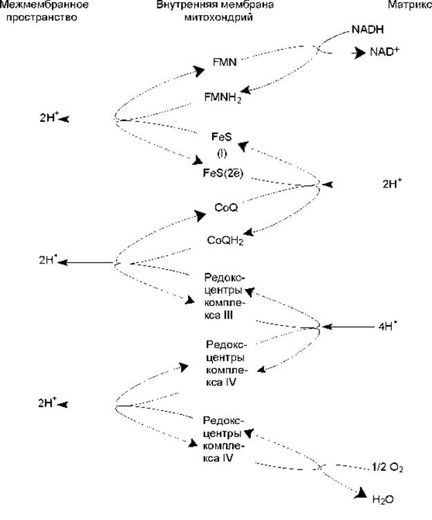
Fig. 12.2. Simplified scheme of electron transport through the Components of the respiratory chain in the inner mitochondrial membrane. Complex I comprises 25—30 subunits, FMN, 6—7 iron-sulfur centers, and is designated as NADH dehydrogenase or NADH—CoQ reductase.
This complex oxidizes NADH and reduces Quinones (CoQ). A hydride ion from NADH and a proton from the mitochondrial matrix are transferred to FMN, which is reduced to FMNH2. FMNH2 carries out The transport of two hydrogen atoms from the inner surface of the membrane to the outer. In the process, two protons are extruded into the intermembrane space, while two electrons are accepted by iron-sulfur Proteins, migrating via their redox centers to the inner surface of the membrane where they are transferred to quinones. However, the reduction of quinones requires hydrogen atoms rather than electrons; therefore, an additional 2 protons are accepted from the matrix (Fig. 12.2).
Next, complex III (CoQH2—cytochrome c reductase) comes into play, containing 11 subunits, 2 iron-sulfur centers, 2 cytochrome b Hemes, and a cytochrome c1 heme. The action of this complex results in The oxidation of CoQH2 and the reduction of cytochrome c. Electron transfer proceeds in the following sequence:
CoQH2 → heme b → FeS (III) → heme c1 → heme c
The oxidation of CoQH2 is also accompanied by the extrusion of protons into the intermembrane space (Fig. 12.2).
Cytochrome c is oxidized by complex IV (cytochrome c oxidase), which includes 13 subunits, 2 copper atoms, 2 heme a groups, and 2 heme a3 groups. This complex reduces molecular oxygen to Water. An electron removed from heme c is transferred to the CuA redox center of cytochrome c oxidase, which is located closest to The surface of the enzyme facing the intermembrane space. Moving further into the membrane interior, the electron is transferred to heme a and then to the heme a3—CuB complex, which ultimately reduces molecular oxygen. It should be noted that while Cytochromes act as electron carriers, cytochrome c oxidase possesses proton-conducting channels and is believed to function as a true proton pump. For every transported electron, 2 protons are translocated: one participates in the reduction of O2, while the second crosses the membrane. Thus, for every 2 electrons transported along the respiratory chain components, 4 protons must be translocated through the cytochrome c oxidase channel (Fig. 12.2).
Complex II (succinate dehydrogenase or succinate-CoQ reductase) transfers hydrogen from succinate—oxidized in the TCA cycle—to quinones without the participation of NAD+. In this case, one fewer 'loop' is formed, and a smaller number of Protons are pumped into the mitochondrial intermembrane space.
Consequently, an electrochemical H+ potential gradient is established across the inner mitochondrial membrane, serving as the driving force for ATP synthesis.
The question of how the energy of the proton gradient is coupled with ATP synthesis remains open. It is known that in intact Cell/35.html">Mitochondria, only ATP synthase permits the reverse movement (down the electrochemical gradient) of protons back into the matrix. This proton flux, obeying the laws of Facilitated Diffusion, occurs through the ATP synthase channel and is somehow coupled with ADP phosphorylation.
ATP synthase (also referred to as ATPase, as under certain conditions it can catalyze the reverse reaction: ATP Hydrolysis and the generation of a proton gradient across the membrane) consists of two parts: a membrane-spanning proton channel (F0) and a 'headpiece' (F1) protruding into the matrix as a mushroom-like appendage. Both parts of ATP synthase, in turn, consist of multiple subunits and are arranged in the membrane in a specific manner (Fig. 12.3). The proton channel and the 'stalk' of the headpiece adjoin each other, and the catalytic centers of the enzyme are located between three α- and three β-subunits.
ATP synthesis takes place in three phases across three catalytic centers. First, ATP synthase binds ADP and Pi; next, a phosphoanhydride bond is formed between them (producing ATP); and finally, ATP is released into the matrix.
It is hypothesized that the energy of protons transported through the F0 channel is expended on the Rotation of the γ-subunit, which alters the conformation of the α- and β-subunits and shifts their catalytic centers into an active state. Other hypotheses explaining the Mechanism of ATP synthesis also exist. According to one of them, proposed by P. Mitchell, a phosphate group (Pi) binds at the active center of the enzyme headpiece in close proximity to the proton channel. The translocation of protons through the F0 channel driven by the pH gradient and Membrane Potential is accompanied by the activation of inorganic phosphate, resulting in the cleavage of a hydroxyl group (which combines with a proton to form a water molecule). The remaining moiety of inorganic phosphate is converted into a highly reactive species that reacts with ADP to form ATP.

Fig. 12.3. Structure of ATP synthase
It should be noted that the proton gradient across Introduction/36.html">Biological Membranes can be utilized not only for ATP synthesis but also for other purposes: the Transmembrane Transport of nucleoside diphosphates and triphosphates, the rotation of bacterial flagella, the maintenance of osmotic pressure, the active Transport of substances against a concentration gradient, and thermogenesis. The latter mechanism is of critical importance for warm-blooded animals, as well as certain plants and insects. Many animals possess a specialized tissue known as 'brown adipose tissue', which contains A large number of mitochondria (the membranes of these mitochondria contain reddish cytochromes). A distinctive feature of such specialized mitochondria is that they utilize the Free energy of the proton gradient not for ATP synthesis, but for thermogenesis: the energy is dissipated as heat.
Last update: 06/08/2026
Editorial and Educational Adaptation: This material has been compiled based on the primary/original source text. The project team performed an editorial review, corrected technical inaccuracies, structured sections, and adapted the content for an educational format.
What was processed:
- elimination of formatting defects (OCR errors, structural breaks, corrupted characters);
- editorial organization of content;
- standardization of terminology in accordance with academic sources;
- verification of factual statements against the original source text.
All mentions of the author, publication year, and origin of the primary text have been preserved in accordance with the source.