Biological Chemistry - Berezov T. T., Korovkin B. F. 1998
Liver
The Role of the Liver in Protein Metabolism
The Role of the Liver in Pigment Metabolism
Let us consider only hemochromogenic pigments, which are formed in the body during The breakdown of Hemoglobin (and, to a significantly lesser extent, during the breakdown of Myoglobin, Cytochromes, etc.). The breakdown of hemoglobin occurs in macrophage Cells, particularly in fixed reticuloendothelial cells (stellate reticuloendotheliocytes) and in histiocytes of the Connective Tissue in any organ.
As noted earlier (see Chapter 13), the initial stage of hemoglobin breakdown is the Cleavage of a single methene bridge, resulting in The formation of verdoglobin. Subsequently, the iron atom and the globin protein are split off from the verdoglobin molecule. This yields biliverdin, which represents a chain of four pyrrole rings linked by methene bridges. Biliverdin is then reduced to bilirubin—a pigment excreted in Bile and therefore referred to as a bile pigment. The resulting bilirubin is designated as indirect (unconjugated) bilirubin. It is insoluble in Water and gives an indirect reaction with the diazo reagent, meaning the reaction occurs only after preliminary Treatment with alcohol.
In the Liver, bilirubin binds (is conjugated) with glucuronic acid. This reaction is catalyzed by the enzyme UDP-glucuronosyltransferase, with glucuronic acid entering the reaction in its active form, i.e., as UDPGA. The resulting bilirubin glucuronide is known as direct bilirubin (conjugated bilirubin). It is soluble in water and yields a direct reaction with the diazo reagent. The greater part of bilirubin combines with two molecules of glucuronic acid, forming bilirubin diglucuronide:
Class="center">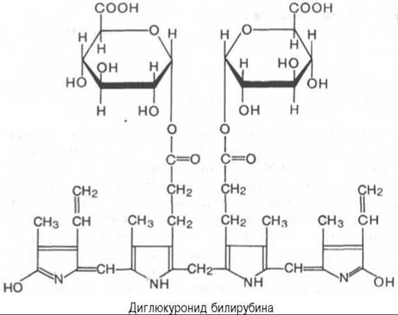
* Written in co-authorship with Candidate of Medical Sciences P.P. Minin.

Fig. 16.4. Normal METABOLISM of urobilinogen bodies (scheme).
Direct bilirubin formed in the liver, along with a very small fraction of indirect bilirubin, is excreted via bile into the Small Intestine. Here, glucuronic acid is cleaved from direct bilirubin, followed by its reduction with the sequential formation of mesobilirubin and mesobilirubinogen (urobilinogen). It is generally accepted that about 10% of bilirubin is reduced to mesobilirubinogen on its way to the small intestine, i.e., within the extrahepatic biliary tract and the Gallbladder. From the small intestine, a portion of the formed mesobilirubinogen (urobilinogen) is reabsorbed through the intestinal wall, enters the portal vein, and is carried by the bloodstream to the liver, where it is completely broken down into di- and tripyrroles. Thus, under normal conditions, mesobilirubinogen does not enter the general Blood Circulation or the urine.
The bulk of mesobilirubinogen passes from the small intestine into the Large Intestine, where it is reduced to stercobilinogen by anaerobic microflora. In the lower sections of the large intestine (mainly in the rectum), the resulting stercobilinogen is oxidized to stercobilin and excreted with feces. Only a minor fraction of stercobilinogen is absorbed into the INFERIOR VENA CAVA system (initially entering the hemorrhoidal Veins) and is subsequently excreted in the urine. Consequently, under normal conditions, human urine contains only traces of stercobilinogen (up to 4 mg per day is excreted in urine). Unfortunately, until recently in clinical practice, stercobilinogen present in normal urine continues to be termed urobilinogen. Fig. 16.4 schematically illustrates the pathways of urobilinogen body formation in The Human Body.
In clinical practice, the term "urine urobilinogen" has become firmly established. This term refers to those derivatives of bilirubin (bilirubinoids) that are detected in urine. A positive reaction for urobilinogen may be caused by an elevated level of a specific bilirubinoid in the urine and generally reflects an underlying pathology.
The clinical determination of blood bilirubin levels (total, indirect, and direct), as well as urine urobilinogen, is of paramount importance in the Cytology/practical/136.html">Differential Diagnosis OF jaundice of various etiologies (Fig. 16.5). In hemolytic jaundice ("suprahepatic"), enhanced hemolysis of erythrocytes and the destruction of hemoglobin lead to the intensive formation of indirect bilirubin within the reticuloendothelial system (see Fig. 16.5, b). The liver proves incapable of utilizing such a massive amount of indirect bilirubin, resulting in its accumulation in the blood and Tissues. Concurrently, the liver synthesizes elevated amounts of direct bilirubin, which enters the intestine with bile. In the small intestine, increased quantities of mesobilirubinogen and subsequently stercobilinogen are formed. The reabsorbed fraction of mesobilirubinogen is utilized by the liver, whereas stercobilinogen reabsorbed in the large intestine is excreted in the urine. Thus, hemolytic jaundice is typically characterized by the following clinical and laboratory findings: elevated levels of total and indirect bilirubin in the blood; absence of bilirubin in the urine (indirect bilirubin is not filtered by the Kidneys) and a positive urobilinogen reaction (due to increased entry of stercobilinogen, and in severe cases mesobilirubinogen not utilized by the liver, into the blood and urine); lemon-yellow Skin tint (a combination of jaundice and anemia); Splenomegaly; and brightly pigmented feces.
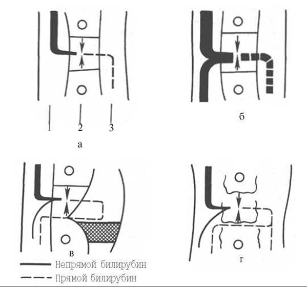
Fig. 16.5. Pathogenesis of bilirubinemia in various pathological states (scheme).
a - normal; b - hemolysis; c - stasis in bile capillaries; d - damage to hepatic parenchymal cells; 1 - blood capillary; 2 - liver cells; 3 - bile capillary.
In obstructive (mechanical, or "subhepatic") jaundice (see Fig. 16.5, c), bile outflow is impaired (obstruction of the common bile duct by a gallstone, carcinoma of the HEAD of the Pancreas). This leads to destructive Changes in the liver and The entry of bile components (bilirubin, Cholesterol, bile acids) into the blood. With complete obstruction of the common bile duct, bile fails to reach the intestine; consequently, no bilirubinoids are formed in the gut, stools are acholic (clay-colored), and the urine urobilinogen reaction is negative. Thus, in obstructive jaundice, blood levels of total bilirubin (predominantly direct) are elevated, along with increased Cholesterol and Bile acids, while urine exhibits high levels of direct bilirubin. Clinical Features of obstructive jaundice include intense yellow skin coloration, pale stools, and pruritus (caused by the irritation of nerve endings by bile acids deposited in the skin). It should be noted that prolonged mechanical jaundice can significantly impair liver Functions, including its primary detoxification function. In such cases, the liver may partially "refuse" indirect bilirubin, leading to its accumulation in the blood. In other words, an elevated indirect bilirubin fraction in obstructive jaundice is an unfavorable prognostic sign.
In parenchymal ("hepatic") jaundice (see Fig. 16.5, d), which most commonly arises from viral infections, inflammatory and destructive processes develop in the liver, leading to impaired liver function. In the Early stages of hepatitis, the uptake and glucuronidation of indirect bilirubin are preserved; however, due to the destruction of the hepatic parenchyma, part of the newly formed direct bilirubin enters the systemic circulation, causing jaundice. Bile excretion is also impaired, and less bilirubin reaches the intestine than under normal conditions. Consequently, less mesobilirubinogen is formed, and a smaller amount is absorbed in the gut. Yet, even this small amount reaching the liver is not utilized by it. Mesobilirubinogen "escapes" into the blood and is subsequently excreted in the urine, resulting in a positive urobilinogen reaction. The amount of stercobilinogen formed is likewise reduced, leading to hypocholic feces. Thus, parenchymal jaundice is marked by elevated blood concentrations of total bilirubin, predominantly at the expense of the direct fraction. Fecal stercobilinogen content is decreased. The urine urobilinogen reaction is positive due to the appearance of mesobilirubinogen in the urine. It should be pointed out that in progressive hepatitis, when the liver loses its detoxification capacity, significant amounts of indirect bilirubin also accumulate in the blood. Furthermore, in cases of severe inflammation and Swelling of the liver, compression of bile capillaries and ductules may occur, precipitating intrahepatic cholestasis, which imparts features of mechanical jaundice to parenchymal jaundice, complete with corresponding clinical and laboratory findings (acholic stools, negative urobilinogen reaction).
Table 16.2 outlines the most characteristic shifts in clinical and laboratory parameters across various types of jaundice.
It should be borne in mind that in clinical practice, a "pure" form of a single type of jaundice is rarely observed. Combinations of different types are much more common. For instance, severe hemolysis inevitably affects various Organs, including the liver, which may introduce elements of parenchymal jaundice into hemolytic conditions. Conversely, parenchymal jaundice generally incorporates elements of mechanical obstruction. In mechanical jaundice resulting from the compression of the major duodenal papilla (ampulla of Vater) by a pancreatic head carcinoma, heightened hemolysis is an inevitable consequence of neoplastic intoxication.
Table 16.2. Differential diagnosis of various types of jaundice

Designations: N - normal; ↑ increased; ↓ decreased; + detected; 0 not detected.
Last update: 06/08/2026
Editorial and Educational Adaptation: This material has been compiled based on the primary/original source text. The project team performed an editorial review, corrected technical inaccuracies, structured sections, and adapted the content for an educational format.
What was processed:
- elimination of formatting defects (OCR errors, structural breaks, corrupted characters);
- editorial organization of content;
- standardization of terminology in accordance with academic sources;
- verification of factual statements against the original source text.
All mentions of the author, publication year, and origin of the primary text have been preserved in accordance with the source.