Biological Chemistry - Berezov, T. T., Korovkin, B. F. 1998
Metabolism of Simple Proteins
Specific Pathways of Amino Acid Metabolism
Metabolism of Dicarboxylic Amino Acids
The classical works of Soviet scientists A.E. Braunstein and S.R. Mardashev and American biochemist A. Meister proved the crucial role of dicarboxylic Amino Acids (glutamic and aspartic acids and their amides, glutamine and asparagine) in integrating Nitrogen METABOLISM within the Organism. The dicarboxylic amino acid system, which also includes the corresponding a-keto acids, is closely tied not only to nitrogen Metabolism as a whole, but also to lipid and Carbohydrate Metabolism. Previous studies highlighted the specific role of dicarboxylic Amino Acids and the Enzymes catalyzing their transformations in redistributing nitrogen in the body, deamination, and synthesizing Natural Amino Acids (transdeamination and transreamination reactions), as well as forming the End products of Protein metabolism, namely urea synthesis.
The main Catabolic pathways for The conversion of dicarboxylic amino acids and their amides can be represented by the following reactions:
Class="center">
Aspartic acid is directly involved in the Ornithine Urea Cycle, Transamination reactions, and The Biosynthesis of CARBOHYDRATES (as a glycogenic amino acid), carnosine, anserine, purine, and pyrimidine NUCLEOTIDES (see Chapter 14), as well as the Synthesis of N-acetylaspartic acid in Brain tissue. The function of the latter, found in relatively high concentrations in mammalian brain tissue, remains to be elucidated.
Glutamic acid, a glycogenic and non-essential amino acid for humans and animals, is also incorporated into the synthesis of several specific metabolites, notably Glutathione and glutamine. Besides participating in Ammonia Transport and acid-base regulation, glutamine serves as an indispensable nitrogen source in numerous synthetic pathways, such as the Biosynthesis of Purine and pyrimidine nucleotides and aminosugars, the detoxification of phenylacetic acid (phenylacetylglutamine synthesis) in humans and anthropoid apes, and the Synthesis of the vitamin Folic acid (pteroylglutamic acid). Figure 12.8 summarizes the synthetic reactions of various substances in which the amide nitrogen of glutamine plays a specific role that cannot be fulfilled by the nitrogen of Other Amino Acids*.

Fig. 12.8. Utilization of glutamine amide nitrogen for the synthesis of various compounds in living organisms.
Furthermore, glutamine and asparagine have proven to be essential growth factors for certain normal and tumor Cells in tissue culture; they cannot be replaced by each other or by the corresponding dicarboxylic amino acids. This indicates that when cultured in vitro, certain cells lose The ability to synthesize these amides via synthetase or transaminase pathways.
Research in Meister's laboratory provided evidence that, in animal Tissues, glutamine and asparagine undergo coupled transamination and deamidation mediated by specific amide transaminases (glutamine transaminase and asparagine transaminase) and nonspecific w-amidase:
* A specific class of enzymes known as glutamine amidotransferases participates in these synthetase reactions. They contain a glutamine-binding domain with a conserved Amino Acid Sequence and an acceptor domain with a variable region for binding the second substrate. Mechanistically, these enzymes are similar to glutaminase (see below), although the latter does not require a second substrate.
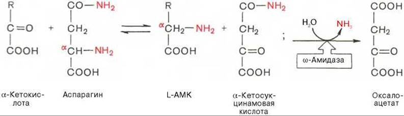
Thus, it is the a-amino group of asparagine rather than the amide group—as previously assumed—that participates in the transfer reaction; meanwhile, the amide group of the intermediate compound, a-ketosuccinamic acid, is subsequently released via Hydrolysis as ammonia. Because transamination is a reversible process, the limiting factors in asparagine (and glutamine) synthesis are the w-amides of oxaloacetate and a-ketoglutaric acid, the synthesis of which in animal tissues has not yet been demonstrated.
Along with glucose, glutamic acid is one of the few compounds that serves as an energy source for brain tissue. Previous studies noted the high activity in brain tissue of glutamate decarboxylase, which catalyzes the conversion of glutamate to y-aminobutyric acid (GABA). Subsequent sequential oxidation of GABA involves transamination to yield succinic semialdehyde, oxidation to succinic acid, and finally oxidation via The Citric Acid Cycle.
Both reactions (glutamate decarboxylase and GABA transamination) require Pyridoxal phosphate, which was found to be more firmly bound to GABA transaminase. GABA exerts an inhibitory effect on synaptic transmission in the Central Nervous system (CNS); consequently, the convulsions observed during vitamin B6 deficiency may be linked to reduced GABA production in the glutamate decarboxylase reaction. In animals, seizures can also be triggered by administering isoniazid, which binds the aldehyde group of the coenzyme, or vitamin B6 antagonists such as methoxypyridoxine. Since GABA is a naturally occurring tranquilizer, one way to increase its concentration in the CNS is to administer agents that inhibit GABA transaminase, which effectively breaks down GABA.
In recent years, an entirely novel pathway for synthesizing glutamic acid from a-ketoglutaric acid and glutamine has been discovered in Bacteria and plants (though not in animal tissues). Termed the glutamate synthase cycle, this pathway involves two irreversible reactions coupled with ATP breakdown, leading to the assimilation of ammonia:
a) NH3 + Glutamate + ATP —> Glutamine + ADP + Pi
b) Glutamine + a-Ketoglutarate + NADPH + H+ —> 2Glutamate + NADP+
NH3 + a-Ketoglutarate + NADPH + H+ + ATP —> Glutamate + NADP+ + ADP + Pi
The First stage (a) is catalyzed by Glutamine Synthetase, found in animal cells, while the Second Stage (b) is catalyzed by glutamate synthase, discovered exclusively in plants, Fungi, and microorganisms. Both stages, together with the reversibly operating Glutamate dehydrogenase reaction (c), can be represented by the following scheme:

It turns out that at low ammonia concentrations characteristic of plants and microorganisms, reactions proceed predominantly via the glutamate synthase cycle, whereas at high concentrations typical of animal tissues, they follow the glutamate dehydrogenase pathway; glutamate is synthesized in both cases.
The summary scheme outlines the main integrative pathways for the conversion of glutamine and glutamic acid and lists the enzymes catalyzing these reactions in tissues (Fig. 12.9).
The Metabolic pathways of Proline and Arginine are also linked to glutamic acid metabolism (see Fig. 12.9), although it should be recalled that arginine is a semi-essential amino acid for the organism, particularly during youth when its synthesis from glutamate cannot meet the demands of rapid growth. The primary metabolic pathway for arginine is urea synthesis. More specific and irreversible is the pathway converting Histidine (also a semi-essential amino acid for animals) into glutamic acid. This transformation involves two well-characterized enzymes: histidine ammonia-lyase (histidase), which catalyzes the intramolecular deamination of histidine, and Urocanase, which catalyzes the Cleavage of the imidazole ring of urocanic acid to form imidazolylpropionic acid; the latter is converted to glutamic acid via formiminoglutamate. Other pathways of Histidine Metabolism (histamine formation and its oxidation by diamine oxidase) were discussed earlier.

Fig. 12.9. Metabolic transformations of glutamate and glutamine in animal tissues (based on Meister's scheme).
1 - citric acid cycle reactions; 2 - glutamate dehydrogenase; 3 - glutamate transaminase; 4 - glutamine synthetase; 5 - glutaminase; 6 - glutamine transaminase; 7 - carbamoyl phosphate synthetase (Liver); 8 - ω-amidase; 9 - γ-glutamylcysteine synthetase; 10 - glutathione synthetase; 11 - γ-glutamyltransferase; 12 - γ-glutamylcyclotransferase; 13 - 5-oxoprolinase; 14 - cysteinyl-glycinase; 15 - glutamate decarboxylase; 16 - glutamate-N-acetylase; 17 - enzymes catalyzing the degradation of these amino acids; 18 - glutamine amidotransferases; 19 - glutamine-phenylacetyltransferase.
Last update: 06/08/2026
Editorial and Educational Adaptation: This material has been compiled based on the primary/original source text. The project team performed an editorial review, corrected technical inaccuracies, structured sections, and adapted the content for an educational format.
What was processed:
- elimination of formatting defects (OCR errors, structural breaks, corrupted characters);
- editorial organization of content;
- standardization of terminology in accordance with academic sources;
- verification of factual statements against the original source text.
All mentions of the author, publication year, and origin of the primary text have been preserved in accordance with the source.