Biological Chemistry - Berezov T. T., Korovkin B. F. 1998
Carbohydrate Metabolism
Gluconeogenesis
Gluconeogenesis is the Synthesis of glucose from non-carbohydrate precursors. Such precursors or metabolites primarily include lactic and pyruvic acids, the so-called glycogenic Amino Acids, glycerol, and several Other Compounds. In other words, the precursors of glucose in gluconeogenesis can be Pyruvate or any compound that is converted into pyruvate or one of the intermediates of The Tricarboxylic Acid Cycle during Catabolism*.
In vertebrates, gluconeogenesis occurs most intensively in the Cells of The Liver and Kidneys (in the renal cortex).
Most steps of gluconeogenesis represent the reversal of Glycolysis. Only 3 reactions of glycolysis (the hexokinase, Phosphofructokinase, and pyruvate kinase reactions) are irreversible, which is why Other Enzymes are employed at these 3 stages in gluconeogenesis. Let us examine The pathway of glucose synthesis from pyruvate.
* In Higher Plants and microorganisms, The Glyoxylate cycle plays a vital role in gluconeogenesis. Thanks to this cycle, higher plants and microorganisms are able to convert two-carbon metabolites, and consequently acetyl-CoA, into CARBOHYDRATES. Animal cells lack two Key Enzymes of the glyoxylate cycle: isocitrate lyase and malate synthase, which is why this cycle cannot operate in them.
Formation of phosphoenolpyruvate from pyruvate. The synthesis of phosphoenolpyruvate is carried out in several stages. Initially, pyruvate is carboxylated* under METABOLISM/18.html">The Influence of pyruvate carboxylase with the participation of CO2 and ATP to form oxaloacetate:
Class="center">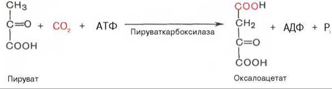
Then, oxaloacetate is converted into phosphoenolpyruvate via decarboxylation and phosphorylation catalyzed by the enzyme phosphoenolpyruvate carboxykinase. Guanosine triphosphate (GTP) serves as the donor of the phosphate group in this reaction:
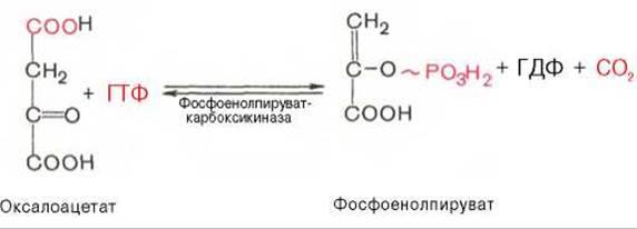
It has been established that enzymes of both the Cytosol and Cell/35.html">Mitochondria participate in The formation of phosphoenolpyruvate.
The First stage of synthesis takes place in the mitochondria (Fig. 10.6). Pyruvate carboxylase, which catalyzes this reaction, is an allosteric mitochondrial enzyme. Acetyl-CoA is required as an allosteric activator of this enzyme. The mitochondrial membrane is impermeable to the resulting oxaloacetate. The latter is reduced to malate right there in the mitochondria:
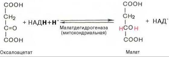
The reaction proceeds with the participation of mitochondrial NAD-dependent malate dehydrogenase. In mitochondria, the NADH/NAD+ ratio is relatively high, due to which intra-mitochondrial oxaloacetate is easily reduced to malate, which readily exits the mitochondrion through the mitochondrial membrane. In the cytosol, the NADH/NAD+ ratio is very low, and malate is oxidized back with the participation of cytoplasmic NAD-dependent malate dehydrogenase:
* The reaction involves the so-called active form of CO2, the formation of which, In addition to ATP, involves biotin (see Chapter 7).
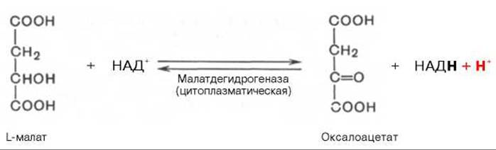
Further conversion of oxaloacetate into phosphoenolpyruvate occurs in The Cell cytosol.
Conversion of fructose-1,6-bisphosphate to fructose-6-phosphate. The phosphoenolpyruvate formed from pyruvate is converted into fructose-1,6-bisphosphate via a series of reversible glycolysis reactions. This is followed by the phosphofructokinase reaction, which is irreversible. Gluconeogenesis bypasses this endergonic reaction. The conversion of fructose-1,6-bisphosphate to fructose-6-phosphate is catalyzed by a specific phosphatase:

Formation of glucose from glucose-6-phosphate. In the subsequent reversible stage of glucose Biosynthesis, fructose-6-phosphate is converted into glucose-6-phosphate. The latter can be dephosphorylated (i.e., bypassing the hexokinase reaction) under the Influence of the enzyme glucose-6-phosphatase:


Fig. 10.6. Formation of phosphoenolpyruvate from pyruvate.
1 - pyruvate carboxylase; 2 - malate dehydrogenase (mitochondrial); 3 - malate dehydrogenase (cytoplasmic); 4 - phosphoenolpyruvate carboxykinase.
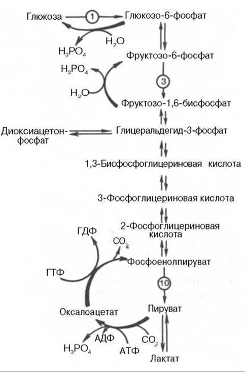
Fig. 10.7. Glycolysis and gluconeogenesis. The red arrows indicate the "bypass" pathways of gluconeogenesis during the BIOSYNTHESIS OF GLUCOSE from pyruvate and lactate; the numbers in circles denote the corresponding stage of glycolysis.
Fig. 10.7 illustrates the "bypass" reactions of gluconeogenesis during The biosynthesis of glucose from pyruvate and lactate.
Regulation of gluconeogenesis. An essential checkpoint in The regulation of gluconeogenesis is the reaction catalyzed by pyruvate carboxylase. Acetyl-CoA acts as a positive allosteric modulator of this enzyme. In the absence of acetyl-CoA, the enzyme is almost completely devoid of catalytic activity. When mitochondrial acetyl-CoA accumulates within the cell, the biosynthesis of glucose from pyruvate is accelerated. Notably, acetyl-CoA simultaneously Functions as a negative modulator of the pyruvate dehydrogenase complex (see below). Consequently, the accumulation of acetyl-CoA slows down The oxidative decarboxylation of pyruvate, thereby further directing the latter toward gluconeogenesis.
Another critical point in the regulation of gluconeogenesis is the reaction catalyzed by fructose-1,6-bisphosphatase, an enzyme inhibited by AMP. AMP exerts the opposite effect on phosphofructokinase, serving as an allosteric activator for this enzyme. When AMP concentrations are low and ATP levels are high, gluconeogenesis is stimulated. Conversely, when the ATP/AMP ratio is low, glucose breakdown (glycolysis) predominates in the cell.
In 1980, a group of Belgian researchers (H. Hers et al.) discovered fructose-2,6-bisphosphate in liver tissue, which acts as a potent regulator of the activities of both aforementioned enzymes:
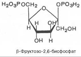
Fructose-2,6-bisphosphate activates phosphofructokinase and inhibits fructose-1,6-bisphosphatase. An increase in the intracellular level of fructose-2,6-bisphosphate enhances glycolysis while suppressing The rate of gluconeogenesis. Conversely, a decrease in fructose-2,6-bisphosphate concentration yields the opposite effect.
It has been established that fructose-2,6-bisphosphate is synthesized from fructose-6-phosphate in an ATP-dependent reaction, and is degraded back into fructose-6-phosphate and inorganic phosphate. Both the Biosynthesis and the degradation of fructose-2,6-bisphosphate are catalyzed by a single bifunctional enzyme exhibiting both phosphokinase and phosphatase activities:
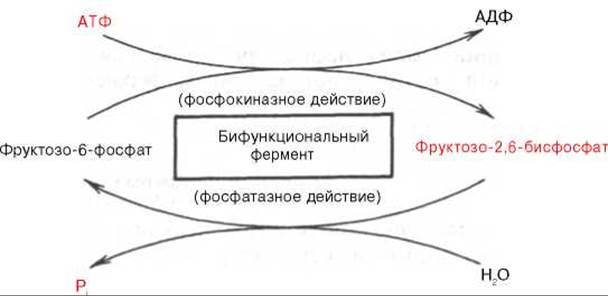
It has also been demonstrated that this bifunctional enzyme is, in turn, regulated via cAMP-dependent phosphorylation. Phosphorylation leads to an increase in phosphatase activity and a decrease in phosphokinase activity of the bifunctional enzyme. This mechanism explains the rapid hormonal response—particularly that of Glucagon—on intracellular fructose-2,6-bisphosphate levels (see Chapter 16).
The activity of the bifunctional enzyme is also modulated by certain metabolites, among which glycerol-3-phosphate is of primary importance. The Effect of glycerol-3-phosphate on the enzyme parallels the effect observed upon its phosphorylation by cAMP-dependent protein Kinases.
Currently, fructose-2,6-bisphosphate has been detected not only in the liver, but also in various other animal Organs and Tissues, as well as in plants and microorganisms.
Research has shown that gluconeogenesis can also be regulated indirectly, via alterations in the activity of enzymes not directly involved in glucose synthesis. For instance, the glycolytic enzyme pyruvate kinase exists in two forms: L and M. The L-form (derived from 'liver') predominates in tissues capable of gluconeogenesis. This form is inhibited by excess ATP and Certain amino acids, notably Alanine. The M-form (derived from 'Muscle') is not subject to this type of regulation. Under conditions of abundant cellular energy, the L-form of pyruvate kinase is inhibited, which slows down glycolysis and creates favorable conditions for gluconeogenesis.
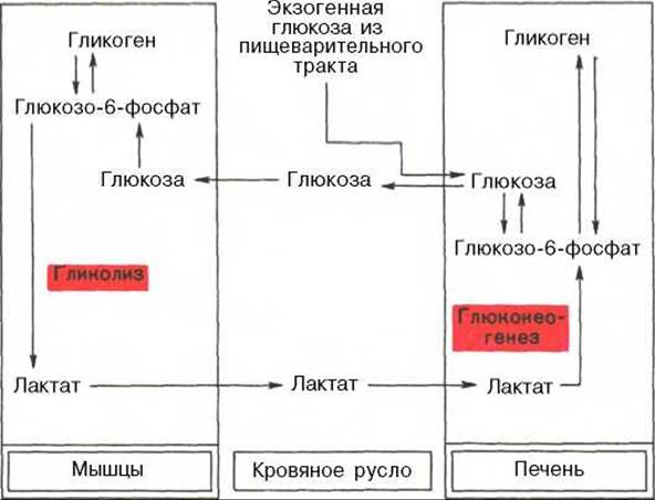
Finally, It is interesting to note the close interplay between glycolysis, which proceeds intensively in Skeletal Muscle during high activity, and gluconeogenesis, which is particularly characteristic of the liver. During maximal muscle exertion, enhanced glycolysis produces an excess of lactic acid that diffuses into the bloodstream; a significant portion of this lactate is then converted into glucose in the liver via gluconeogenesis. This glucose can subsequently serve as an energy substrate required for muscle performance. The metabolic coupling between glycolysis in Muscle tissue and gluconeogenesis in the liver can be represented by the following scheme:
Last update: 06/08/2026
Editorial and Educational Adaptation: This material has been compiled based on the primary/original source text. The project team performed an editorial review, corrected technical inaccuracies, structured sections, and adapted the content for an educational format.
What was processed:
- elimination of formatting defects (OCR errors, structural breaks, corrupted characters);
- editorial organization of content;
- standardization of terminology in accordance with academic sources;
- verification of factual statements against the original source text.
All mentions of the author, publication year, and origin of the primary text have been preserved in accordance with the source.