Biochemistry and Molecular Biology - Belyasova N.A. 2002
Molecular Foundations and Mechanisms of Heredity
Organization of the Cellular Genetic Apparatus
Types of Nucleic Acids and Their Functions
Of the two Types of Nucleic acids—DNA and RNA—deoxyribonucleic acid serves as the substance that stores all the primary Genetic information of The Cell and is capable of self-Replication, whereas Ribonucleic Acids act as intermediaries between DNA and protein. These Functions of Nucleic Acids are closely related to the Specific features of their individual structures.
DNA and RNA are polymeric macromolecules whose monomers are NUCLEOTIDES. Each nucleotide consists of three components: a monosaccharide, a phosphoric acid residue, and a nitrogenous base. The nitrogenous base is linked to the sugar via a ß-N-glycosidic bond (Fig. 1.1).
The sugar incorporated into the nucleotide (a pentose) can exist in one of two forms: ß-D-ribose or ß-D-2-deoxyribose. The difference between them is that the hydroxyl group of ribose at the 2'-carbon atom of the pentose is replaced by a hydrogen atom in deoxyribose. Nucleotides containing ribose are called ribonucleotides and serve as the monomers of RNA, whereas nucleotides containing deoxyribose are termed deoxyribonucleotides and form DNA.
Nitrogenous bases are derivatives of one of two parent compounds: purine or pyrimidine. Nucleic acids predominantly contain two purine bases—adenine (A) and guanine (G)—and three pyrimidine bases—cytosine (C), thymine (T), and uracil (U). Ribonucleotides and, accordingly, RNA contain the bases A, G, C, and U, whereas deoxyribonucleotides and DNA contain A, G, C, and T.
Class="center">
Fig. 1.1. Structure of a nucleoside and a nucleotide: the numbers indicate the positions of atoms in the pentose residue
The nomenclature of Nucleosides and Nucleotides is widely used in biochemistry and molecular biology and is summarized in Table 1.1.
Table 1.1. Nomenclature of nucleotides and nucleosides

Long polynucleotide chains of DNA and RNA are formed by linking nucleotides together through phosphodiester bonds. Each phosphate group connects the hydroxyl group at the 3'-carbon atom of the pentose of one nucleotide to the OH group at the 5'-carbon atom of the pentose of the adjacent nucleotide (Fig. 1.2).
Acid Hydrolysis of nucleic acids yields individual nucleotide components, whereas Enzymatic hydrolysis using Nucleases cleaves specific bonds within the phosphodiester backbone, thereby exposing the 3'- and 5'-ends of the molecule (Fig. 1.2).
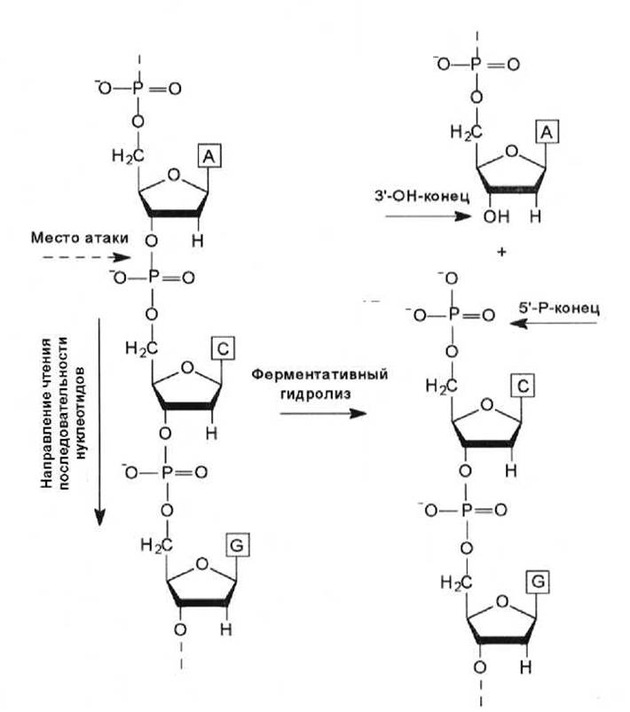
Fig. 1.2. Introduction/11.html">Secondary structure of DNA. Enzymatic hydrolysis of a DNA strand exposing the 3'- and 5'-free ends of the molecule
This provides a basis for considering the nucleic acid strand as polar, making it possible to determine the direction in which The nucleotide sequence is read. It should be noted that most Enzymes involved in the synthesis and hydrolysis of nucleic acids operate in the 5' to 3' direction (5' → 3') of the nucleic acid strand. According to the accepted convention, nucleotide sequences in nucleic acid strands are also read in the 5' → 3' direction (Fig. 1.2).
Structural Features of DNA. According to the three-dimensional model proposed by Watson and Crick in 1953, a DNA molecule consists of two polynucleotide chains that form a right-handed helix around a common axis. The strands within the molecule run in opposite directions and maintain a nearly constant diameter along with other parameters that are independent of nucleotide composition—unlike Proteins, where The sequence of amino acid residues determines the secondary and Tertiary Structure of the molecule.
The sugar-phosphate backbone is located on the periphery of the helix, while the nitrogenous bases are oriented inward with their planes perpendicular to the helical axis. Specific Hydrogen Bonds form between Base Pairs situated opposite each other in the complementary strands: adenine always pairs with thymine, and guanine with cytosine. Specifically, an AT base pair is held together by two hydrogen bonds—one formed between the amino and keto groups, and the other between two nitrogen atoms of the purine and pyrimidine, respectively. A GC base pair features three hydrogen bonds: two formed between the amino and keto groups of the respective bases, and the third between the nitrogen atom of the pyrimidine and the hydrogen (attached to the nitrogen atom) of the purine.
Thus, the larger Purines always pair with the smaller Pyrimidines. As a result, the distances between the C1' atoms of deoxyribose in the two strands are identical for both AT and GC pairs, measuring 1.085 nm. These Two Types of nucleotide pairs, AT and GC, are referred to as complementary pairs. Base pairing between two purines, two pyrimidines, or non-complementary bases (A+C or G+T) is sterically hindered because appropriate hydrogen bonds cannot form, thereby disrupting the geometry of the helix.
The geometry of The Double Helix is such that adjacent nucleotides within a strand are spaced 0.34 nm apart. There are 10 nucleotide pairs per helical turn, and the pitch of the helix is 3.4 nm (10 × 0.34 nm). The diameter of the double helix is approximately 2.0 nm. Because the sugar-phosphate backbone is positioned further from the helical axis than the nitrogenous bases, the double helix features Major and minor grooves (Fig. 1.3).
The DNA molecule is capable of adopting various Conformations, with A-, B-, and Z-forms having been identified. B-DNA is the conventional form found within the cell, in which the planes of the base rings are perpendicular to the double-helical axis. In the A-form of DNA, the planes of the base pairs are tilted by approximately 20° relative to the normal to the axis of the right-handed double helix. Z-DNA is a left-handed helix with 12 nucleotide pairs per turn. The BIOLOGICAL FUNCTIONS OF the A- and Z-forms of DNA remain not fully understood.

Fig. 1.3. Schematic representation of the B-form of the DNA double helix, showing the major and minor grooves. The distance between adjacent base pairs and the pitch of the helix are indicated [M. Singer, P. Berg, 1998]
The Stability of the double helix is maintained by hydrogen bonds between complementary nucleotides in antiparallel strands, stacking interactions (interplanar Van der Waals contacts between atoms and the overlap of $\pi$-orbitals of the contacting bases), and hydrophobic interactions. The latter are manifested by the nonpolar nitrogenous bases being directed toward the interior of the helix, shielding them from direct contact with the polar solvent, whereas the charged sugar-phosphate groups are oriented outward to interact with the solvent.
Because the two DNA strands are held together solely by non-covalent bonds, the DNA molecule readily dissociates into individual strands upon heating or in alkaline solutions (Denaturation). However, upon slow cooling (annealing), the strands are capable of re-associating, and Hydrogen bonds are restored between the complementary bases (renaturation). These properties of DNA are of paramount importance for Genetic Engineering methodology (Chapter 20).
The size of DNA molecules is expressed in terms of the number of nucleotide pairs, with one thousand nucleotide pairs, or 1 kilobase (kb), serving as the unit of measurement. The molecular mass of 1 kb of B-form DNA is ~ 6.6 × 10⁵ Da, and its length is 340 nm. The complete genome of E. coli (~ 4 × 10⁶ bp) is represented by a single circular DNA molecule (nucleoid) and has a length of 1.4 mm.
Structural features and functions of RNA. RNA molecules are single-stranded polynucleotides consisting of 70 to 10,000 nucleotides (sometimes more) and are represented by the following types: mRNA (Messenger RNA), tRNA (Transfer RNA), rRNA (ribosomal RNA), and—exclusively in Eukaryotic Cells—hnRNA (heterogeneous nuclear RNA) and snRNA (small nuclear RNA). These types of RNA perform specific functions; furthermore, in certain Viral Particles, RNA serves as the carrier of genetic information.
Messenger RNA is a transcript of a specific segment of the DNA sense strand and is synthesized during METABOLISM/31.html">Transcription. mRNA serves as a template (matrix) for building a polypeptide molecule. Every three consecutive nucleotides in mRNA function as a codon, determining THE POSITION OF the corresponding amino acid in the peptide. Thus, mRNA acts as an intermediary between DNA and protein.
Transfer RNA also participates in Protein Synthesis. Its function is to deliver Amino Acids to the site of synthesis and determine the position of The amino acid in the peptide. To achieve this, tRNA contains a specific nucleotide triplet called an anticodon, and the entire molecule features a unique structure. The structural model of a tRNA molecule is known as the "cloverleaf" (Fig. 1.4).
The tRNA molecule is short, consisting of 74–90 nucleotides. Like any nucleic acid chain, it has two ends: a phosphorylated 5' end and a 3' end, which always contains three nucleotides—CCA—and a terminal 3'-OH group. An amino acid attaches to the 3' end of the tRNA, which is referred to as the acceptor stem. The tRNA contains several unusually modified nucleotides that are not found in other nucleic acids.

Fig. 1.4. Structure of tRNA [E. Rees, M. Sternberg, 1988]
Although the tRNA molecule is single-stranded, it contains distinct duplex regions that form so-called stems or arms, where Watson-Crick base pairs form between asymmetric PARTS OF THE chain (Fig. 1.4). All known tRNAs form a cloverleaf with four stems (acceptor, D, anticodon, and T). The stems take the form of a right-handed double helix, known as the A-form of DNA. The tRNA loops represent single-stranded regions. Some tRNAs have additional loops and/or stems (e.g., the variable loop of Yeast phenylalanine tRNA).
Recognition of the corresponding site in the mRNA by the tRNA molecule is mediated by the anticodon located in the anticodon loop (Fig. 1.4). Hydrogen bonds form between the bases of the codon and anticodon, provided that the sequences forming them are complementary and the polynucleotide chains are antiparallel (Fig. 1.5).
Molecules of different tRNAs differ in their nucleotide sequences, yet their tertiary structure is highly similar. The molecule folds in such a way that it resembles the letter L in shape. The acceptor and T stems are spatially arranged to form a single continuous helix—the "crossbar" of the L; the anticodon and D stems form the "leg." The proper spatial folding of tRNA molecules is crucial for their function.
Quantitatively, ribosomal RNA predominates in the cell, yet its diversity compared to Other types of RNA is the lowest: rRNA accounts for up to 80% of the mass of cellular RNA and is represented by only three to four types. Meanwhile, the mass of nearly 100 types of tRNA is about 15%, and the proportion of several thousand different mRNAs is less than 5% of cellular RNA mass.
In E. coli cells, 3 types of rRNA are found: 5 S, 16 S, and 23 S, whereas Eukaryotic cells contain 18 S, 5.8 S, 28 S, and 5 S rRNAs. These types of rRNA are components of Ribosomes and make up approximately 65% of their mass. Within ribosomes, rRNAs are densely packed and capable of folding to form base-paired stems similar to those in tRNA. It is believed that rRNAs participate in binding the ribosome to tRNA; specifically, it has been shown that 5 S rRNA interacts with the T-arm of tRNA.
In addition to the aforementioned types of RNA, heterogeneous nuclear RNA and Small nuclear RNAs have been found in the nuclei of eukaryotes. hnRNA accounts for less than 2% of the total cellular RNA. These molecules undergo rapid turnover—for most of them, the half-life does not exceed 10 min. One of the few identified functions of hnRNA is its role as an mRNA precursor. snRNAs are associated with A number of proteins to form so-called small nuclear ribonucleoprotein particles (snRNPs), which carry out RNA splicing (Chapter 3).

Fig. 1.5. Interaction of the mRNA codon with the tRNA anticodon. Dots indicate hydrogen bonds between complementary nucleotides
Last update: 06/08/2026
Editorial and Educational Adaptation: This material has been compiled based on the primary/original source text. The project team performed an editorial review, corrected technical inaccuracies, structured sections, and adapted the content for an educational format.
What was processed:
- elimination of formatting defects (OCR errors, structural breaks, corrupted characters);
- editorial organization of content;
- standardization of terminology in accordance with academic sources;
- verification of factual statements against the original source text.
All mentions of the author, publication year, and origin of the primary text have been preserved in accordance with the source.