IMMUNOLOGY - Roit I. - Mir 2000
Chapter 15. Evolution of the Immune System
LYMPHOMYELOID TISSUES IN LOWER VERTEBRATES
The anatomical basis for the interaction between immunocytes and Antigens is provided by the lymphomyeloid system, which serves as the site of origin and storage for lymphocytes, granulocytes, and other Blood Cells.
Lymphomyeloid Tissues in fish
Hagfish lack both a Thymus and a Spleen. In these cyclostomes, lymphocytes develop within lymphoid tissue located in the region of the gill slits or the gut. Other cyclostomes, such as lampreys, possess a primitive spleen and Bone Marrow-like tissue.
Gnathostome fish lack the bone marrow lymphoid differentiation Lineage, Lymph Nodes, and gut-associated lymphoid tissue (GALT) (Fig. 15.23). However, they have a well-developed thymus and spleen, diffuse GALT, and lymphomyeloid tissue in the Kidneys and Liver (Fig. 15.24). An important feature of fish lymphomyeloid tissue is the presence of numerous melano-macrophage centers in the liver of "primitive" forms (and also in the spleen and kidneys of teleosts) (Fig. 15.25). These centers accumulate pigments such as hemosiderin, ceroid, melanin, and particularly high amounts of lipofuscin. The accumulation of pigments in the macrophage aggregates of fish may be partly related to the high content of Unsaturated Fatty acids, which helps maintain membrane fluidity at low temperatures; such Lipids are especially prone to Lipid Peroxidation and lipofuscin formation.
Class="center">
Fig. 15.23. In fish and amphibians, lymphoid and myeloid tissues are not segregated.

Fig. 15.24. Note that the intestine in sharks and sturgeons is also rich in lymphomyeloid tissue. (Illustration kindly provided by Dr. R. Fänge.)
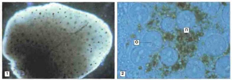
Fig. 15.25. Melano-macrophage centers (MMCs) in the liver of fish. Macro- and microphotographs of the liver of a fish (Rivulus marmoratus) experimentally infected with the parasite Calyptospora funduli. MMCs are clearly visible 60 days post-infection (1, x 60). A coverslip preparation (2, x 100) shows that MMCs consist of degenerating parasite oocytes (O) and adjacent host pigment granules (P). Mononuclear phagocytes play the primary role in MMC formation. (Photographs kindly provided by Dr. W. Vogelbein.)
Lymphomyeloid tissues in amphibians
Thymus. In adult Xenopus, the thymus is located posterior to the Middle ear, directly beneath the Skin. As in most other vertebrates except teleost fish, it separates from the pharyngeal epithelium at very early developmental stages. The thymus differentiates into an outer cortical zone and an inner, more lightly staining medullary zone. Rapidly proliferating lymphocytes of the cortex are particularly sensitive to radiation exposure (Fig. 15.26). In vitro Treatment of the Xenopus thymus with glucocorticoids accelerates Cell apoptosis. In Rana, elevated in vivo corticosteroid levels can induce thymic atrophy.
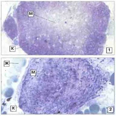
Fig. 15.26. Thymus of an adult Xenopus: effects of irradiation. In a normal (1, x 35) thymus, the cortical zone (C) contains numerous lymphoid elements, whereas the lighter-staining medullary zone (M) is sparsely populated by lymphocytes. The result of gamma irradiation of the thymus (9 days after a 300 rad dose) is shown (2, x 90). Note the dramatic depletion of lymphocytes in the cortex (C) while some cells remain in the medulla (M). The irradiated thymus is reduced in size. The thymus is surrounded by adipose tissue (A). Toluidine blue staining.
There is compelling evidence that, in poikilotherms as well as homeotherms, the thymus produces lymphocytes with T-cell Functions. Figure 15.27 illustrates the ultrastructure of thymic lymphocytes and adjacent epithelial cells. Other stromal cell types are also present in the amphibian thymus, including large dendritic (interdigitating) cells, macrophages, vesicular degenerating cells, and granular cells. Myoid cells (see Fig. 15.27), which may facilitate tissue fluid Circulation and serve as a source of macrophage-stimulating factors, have been identified in the thymus of reptiles and mammals. Thymic epithelial cells expressing MHC class II antigens early in development appear to participate in T-cell "education" (see below). Complexes of stromal cells enclosing thymocytes, reminiscent of nurse cells, are found in the frog thymus and may represent sites of T-cell education. B cells have been detected in the thymus of various vertebrate species, including amphibians, although this organ is not their site of production. High endothelial venules, which may facilitate cell immigration, have been found in the thymus of Xenopus.
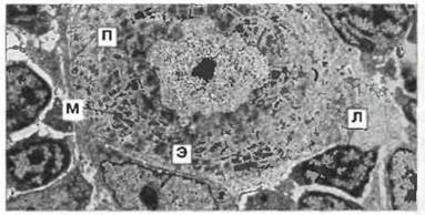
Fig. 15.27. Electron micrograph of the medullary zone of a Xenopus tadpole thymus. The Nucleus of a myoid cell is surrounded by concentric rings of striated myofibrils (M) (resembling Skeletal Muscle). The nuclear Chromatin of small lymphocytes (L) forms distinct electron-dense patches; the Cytoplasm is poor in Organelles. In the epithelial Cell Nucleus (E), chromatin is evenly distributed and nucleoli are visible; the cytoplasm is abundant, and its finger-like projections (P) interdigitate between lymphocytes and other cell types to form a supportive network, x 700. (Photograph kindly provided by Dr. J. Rimmer.)
Spleen. In all gnathostome vertebrates, the spleen functions as the primary peripheral lymphoid organ. Together with LYMPH NODES AND kidneys, it traps antigens, retains lymphocytes that proliferate following antigenic stimulation, and releases these cells and their products into the bloodstream. The Xenopus spleen contains thymus-dependent and thymus-independent lymphoid zones (Fig. 15.28). White pulp follicles contain numerous B cells (Fig. 15.29), which can be visualized by staining this region with Monoclonal Antibodies against IMMUNOGLOBULINS. Splenic T cells, located primarily in the marginal zone, lack surface immunoglobulins but bind anti-T-cell monoclonal antibodies (see Fig. 15.29).

Fig. 15.28. Spleen section from an adult Xenopus. Thymus-dependent [perifollicular red pulp or marginal zone (MZ)] and thymus-independent (white pulp) regions are visible. In Xenopus (unlike many other poikilotherms), the white pulp (WP) is clearly demarcated from the surrounding red pulp (RP) by a poorly staining boundary cell layer (BL). Accumulations of lymphocytes are also visible in the red pulp. Hematoxylin-eosin staining, x 80.

Fig. 15.29. Spleen of an adult Xenopus showing B- and T-cell-rich zones.
1. White pulp (WP) follicle containing numerous B cells; these cells are also present in the marginal zone (MZ) and red pulp (RP), predominantly as intensely stained plasma cells. Anti-B-cell (anti-IgM) mAb staining, x 100.
2. In the marginal zone bordering the white pulp (WP) follicle, clusters of T cells are visible. They are particularly abundant in the perifollicular (marginal) zone (MZ) and lack surface immunoglobulins. Anti-T-cell antibody staining, x 200.
Blood enters the spleen via the central arteriole of the white pulp, which is closely associated with noradrenergic fibers of the sympathetic Nervous system. These fibers innervate the spleen and exert immunomodulatory effects. Capillaries from the central arteriole penetrate the marginal zone surrounding the red pulp. Their walls contribute to The formation of the marginal layer. Experiments using India ink staining and fluorescent antigens have demonstrated that blood-borne material initially enters the red pulp. Later, circulating antigens are trapped by the white pulp follicles and thus come into close contact with potential antibody-producing cells (Fig. 15.30). Antigens are retained On the surface of large dendritic cells, whose processes extend as pseudopodia across the marginal layer into the T-cell-rich marginal zone. The general architecture of the amphibian spleen resembles that of mammals, although it lacks germinal centers. The spleen plays a crucial role in B-cell maturation in amphibians, both during the tadpole stage (along with the liver) and in adults, where it serves as the primary site of B-Cell Differentiation. Surprisingly, Xenopus B lymphocytes constitutively fail to express CD5—a marker present on "primitive" B-I cells in mammals that produce so-called natural antibodies.

Fig. 15.30. Immunofluorescence of an adult Xenopus spleen demonstrating antigen trapping. The frog was injected with human IgG. Three weeks later, frozen spleen sections were prepared and incubated with fluorescein-labeled anti-human IgG antibodies. Bright green fluorescence indicates the presence of antigen in the white pulp (WP) follicles. The antigen is trapped by dendritic cells, which are morphologically similar to those in mammals and birds, and appears on The surface of reticular cells, x 35.
Lymphomyeloid nodes. Lymphomyeloid nodes, presumably analogous in function to the lymph nodes of homoiothermic animals, first appear in vertebrate evolution in "higher" amphibians, such as Rana frogs and Bufo toads, but are absent in urodeles and Xenopus. Unlike their mammalian counterparts, the lymphomyeloid nodes of anuran amphibians function primarily as blood-filtering Organs, although they can also trap material from the surrounding lymph. Serving as a major site for antibody-producing cells, the lymphomyeloid nodes of anurans lack the distinct architecture characteristic of mammalian lymph nodes and do not contain germinal centers. In adult frogs, " lymph nodes" are located in the neck and axillary regions; the lymph glands of tadpoles exhibit a similar Structure (Fig. 15.31).
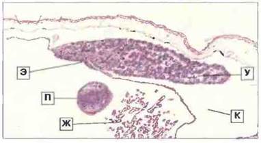
Fig. 15.31. Section of a Rana tadpole lymph gland. The elongated (paired) lymphomyeloid node (N) is attached ventrally to the epithelium (E) of the gill chamber and projects into the lumen of a large lymphatic channel (C). The gills (G) and a forelimb digit (D) lying within the gill chamber are visible in the middle; the tadpole skin is shown at the top. The lymph gland consists of a lymphoid parenchyma containing phagocytes intersected by sinusoids (pale-staining areas). It functions primarily as a blood-filtering organ. Hematoxylin and eosin stain, x 25.
Gut-associated lymphoid tissue. Throughout the length of the amphibian Small Intestine, aggregates of gut-associated lymphoid tissue (GALT) are detected, analogous to this system in mammals. In Xenopus, GALT contains plasma cells that secrete IgM and IgX. Immunohistochemical analysis using anti-T-cell monoclonal antibodies reveals both nodular T-cell aggregates (lamina propria) and intraepithelial T cells in the Xenopus intestine (Fig. 15.32).
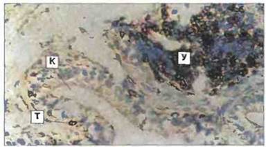
Fig. 15.32. Cryostat section of the Xenopus small intestine stained with anti-CD5 monoclonal antibodies (a pan-T-cell marker) using the immunoperoxidase method. Intraepithelial T cells (T), a nodular T-cell aggregate (N) in the lamina propria, and the intestinal lumen (L) are visible. T cells are absent from both locations in early thymectomized individuals, x 300.
Kidneys and liver. In amphibians, as in fish, the kidneys serve as the primary lymphomyeloid organ (Fig. 15.33), whereas in reptiles, birds, and mammals they lose this function. In anurans, B-cell ontogenetic maturation begins in the kidneys and/or liver. These organs are directly involved in the early differentiation processes of erythroid, lymphoid, and myeloid cells across various vertebrates.
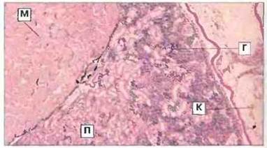
Fig. 15.33. Section of a Rana tadpole Kidney showing a region of hematopoietic tissue. Hematopoietic tissue (H) fills the intertubular spaces and contains lymphocytes, granulocytes, and other maturing blood cells. The mesonephros is adjacent to the myotomal muscle (M) and an intestinal loop (L). Hematoxylin and eosin stain, x 25. (T - renal tubule.)
Bone marrow. Amphibians possess bone marrow, but its immunological role remains unclear. In adult leopard frogs (Rana pipiens), bone marrow lymphomyeloid tissue is readily identified (Fig. 15.34) and serves as an important source of antibody-producing cells. Conversely, in Xenopus, the bone marrow appears to be more rudimentary, serving primarily as a site for neutrophil granulocyte differentiation (Fig. 15.34).
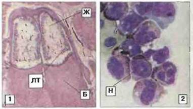
Fig. 15.34. Bone marrow. 1. Lymphomyeloid tissue (LMT), an important source of antibody-producing cells, in the bone marrow of Rana. The Femur (F) and adipose tissue (A) are also visible. Hematoxylin and eosin stain, x 20. 2. Centrifuged bone marrow preparation from Xenopus, showing peroxidase-containing neutrophil granulocytes (N), x 700. (Preparation kindly provided by Dr. I. Hadji-Azimi.)
Last update: 13/08/2026
Editorial and Educational Adaptation: This material has been compiled based on the primary/original source text. The project team performed an editorial review, corrected technical inaccuracies, structured sections, and adapted the content for an educational format.
What was processed:
- elimination of formatting defects (OCR errors, structural breaks, corrupted characters);
- editorial organization of content;
- standardization of terminology in accordance with academic sources;
- verification of factual statements against the original source text.
All mentions of the author, publication year, and origin of the primary text have been preserved in accordance with the source.