General Microbiology - Schlegel H. 1987
Types of fermentation
Formic acid fermentation and the family Enterobacteriaceae
Certain acid-producing microorganisms are grouped into a single physiological category on the basis that formic acid is a characteristic, though not primary, product of their Fermentation. Alongside formic acid, these Bacteria also secrete several other acids; this type of METABOLISM is therefore known as Formic acid fermentation or mixed-acid fermentation. Because some typical members of this group inhabit the intestinal tract, the entire family is named Enterobacteriaceae. They are Gram-negative, actively motile, non-spore-forming rods with peritrichous flagellation. Being facultative anaerobes, they possess Hemoproteins (Cytochromes and catalase) and are capable of generating energy both through Respiration (under aerobic conditions) and fermentation (under anaerobic conditions). Regarding their nutritional requirements, these bacteria are exceptionally undemanding, growing readily on simple synthetic media containing mineral salts, CARBOHYDRATES, and ammonium. Glucose fermentation in all members of this group proceeds with The formation of acids. The Significance of Enterobacteriaceae for Epidemiology as well as for various experimental studies is widely recognized; it is therefore useful to examine some representatives of this family here.
The most important species. Escherichia coli is an intestinal inhabitant, although quantitatively not the dominant one (the intestinal flora is dominated by Bacteroides and Bifidobacterium). This bacterium can maintain viability outside the intestine for some time and is easily detected, a property utilized to monitor drinking Water for fecal contamination.
Class="center">Table 8.4. Characteristics of genera belonging to Enterobacteriaceae
|
Genus |
A plus sign indicates that the majority of strains are positive for the given characteristic |
|||||||
|
Motility |
Glucose fermentation |
Lactose fermentation |
Formation of Н2 |
Indole formation |
Acetoin formation |
Proteolysis |
Urease activity |
|
|
Escherichia |
+ |
+ |
+ |
+ |
+ |
- |
- |
- |
|
Klebsiella |
- |
+ |
+ |
+ |
- |
+ |
- |
+ |
|
Enterobacter |
+ |
+ |
+ |
+ |
- |
+ |
( + ) |
( + ) |
|
Serratia |
+ |
+ |
- |
+ |
- |
+ |
+ |
- |
|
Proteus |
+ |
+ |
- |
+ |
+ |
- |
+ |
+ |
|
Citrobacter |
+ |
+ |
+ |
( + ) |
+ |
- |
- |
( + ) |
|
Salmonella |
+ |
+ |
- |
+ |
- |
- |
- |
- |
|
Shigella |
- |
+ |
- |
- |
+ |
- |
- |
- |
|
Erwinia |
+ |
+ |
( + ) |
- |
( + ) |
( + ) |
_ |
|
Proteus vulgaris is also part of the normal intestinal flora, but is furthermore widespread in soil and water. This bacterium is well known for its pleomorphism (hence its name) and high motility, as well as its ability to form a continuous confluent film on Agar surfaces (Section 2.2.5).
Enterobacter aerogenes can be considered, in a sense, the "twin" of E. coli (both species belong to the "coliform group"). This bacterium is widely distributed in soil; as its species name indicates, it produces abundant gaseous products. It differs from E. coli in only a few biochemical characteristics (see Tables 8.4 and 8.5).
Serratia marcescens (formerly known as Bacterium prodigiosum) can be regarded as a pigmented variant of Enterobacter.
The genus Erwinia includes several phytopathogenic species that affect stems, leaves, and roots. By secreting pectinases, they induce soft rots.
Klebsiella pneumoniae differs from Enterobacter solely by possessing a thicker mucous capsule and being non-motile. It is encountered in certain severe forms of Pneumonia.
Salmonella typhimurium is the most common bacterium causing gastroenteritis, or so-called "food poisoning"; the symptoms stem from irritation of the gastrointestinal mucosa by the bacterium's lipopolysaccharide toxins. The pathogen does not invade the bloodstream. S. typhi is
the CAUSATIVE AGENT OF epidemic typhoid fever, whereas Shigella dysenteriae and related strains cause dysentery.
Vibrio cholerae is the causative agent of cholera, an epidemic disease. It does not belong to the Enterobacteriaceae family, but is closely related to them in its metabolic type. The cholera vibrio multiplies within the intestine, attaching to the intestinal epithelium without penetrating the Cells. Cholera enterotoxin is a protein that binds to specific receptors on intestinal epithelial cells, triggering a net efflux of water, sodium, bicarbonate, and chloride ions into the intestinal lumen.
Yersinia pestis is the causative agent of plague. The genus Yersinia is not taxonomically part of the Enterobacteriaceae, but shares close affinities with them in its facultatively anaerobic lifestyle and fermentation pattern. Wild rodents, primarily rats, serve as the natural reservoir for this epidemic pathogen. The bacteria are transmitted to humans via infected fleas and other ectoparasites, resulting in bubonic or pneumonic plague. Rapid bacterial proliferation within the host and intensive toxin production can lead to rapid death.
Drinking water analysis. The primary objective of drinking water analysis is the detection of Escherichia coli. This analysis serves as a straightforward example of bacteriological Differential Diagnosis and therefore warrants a closer look. E. coli is a common and completely harmless inhabitant of the human intestine, and its presence in drinking water is not hazardous in its own right. However, various pathogenic bacteria may also inhabit the gut. Alongside E. coli, these pathogens are shed in the feces of patients, convalescents, and carriers, and thus may find their way into drinking water. Rather than employing specialized Methods to detect each individual pathogen, a general indicator of contamination is used. E. coli, as a permanent resident of the intestine, serves precisely this indicator role. The detection of this species in a water sample indicates contamination by intestinal contents and enteric bacteria, among which pathogenic forms may also be present. Appropriate measures must be taken under such circumstances. The standard for drinking water stipulates that the total bacterial count per 1 ml must not exceed 100, while 100 ml of water must be entirely free of E. coli cells.
E. coli grows robustly on media containing glucose or lactose and peptone. Lactose is utilized to create conditions that minimize the growth of other bacteria. Only those bacteria capable of cleaving lactose via ß-galactosidase can proliferate on lactose-containing media. This enzyme is synthesized by coliform and lactic acid bacteria, whereas many soil and water species lack it.
Initial indications of gas-producing bacteria are provided by gas evolution during sample incubation in a lactose-peptone solution using Einhorn fermentation tubes. If Escherichia coli is inoculated into one tube and Enterobacter aerogenes into another, a noticeable difference in gas production becomes apparent after just 24 hours of incubation at 37°C. True to its name, E. aerogenes produces roughly twice as much gas as E. coli. The COMPOSITION OF THE evolved gas also differs: E. coli releases H2 and CO2 in an approximate 1:1 ratio, whereas Enterobacter aerogenes generates a higher proportion of CO2 relative to hydrogen.
Table 8.5. Reactions for the differentiation of Escherichia coli and Enterobacter aerogenes
|
Indole production |
Methyl red test |
Acetoin production |
Citrate utilization |
|
|
Escherichia coli |
+ |
+ |
_ |
- |
|
Enterobacter aerogenes |
- |
- |
+ |
+ |
Certain lactic acid bacteria also possess The ability to ferment lactose with gas production, which can potentially confound test results. This necessitates The Use of additional differential techniques. When such a culture is streaked onto eosin methylene blue agar (lactose-peptone-eosin-methylene blue), E. coli forms dark blue colonies with a metallic sheen (resulting from light reflection), whereas Enterobacter produces pink, mucoid colonies lacking a metallic luster.
More precise differentiation of these two microorganisms requires a complete fermentation analysis. While undoubtedly the most accurate method, it is also the most labor-intensive. In routine practice, final differentiation relies on a method based on qualitative differences between the two bacterial species under consideration (Table 8.5). The following parameters are evaluated: 1) indole production from Tryptophan; 2) The amount of acid produced from sugar (methyl red test); 3) acetoin formation during glucose fermentation (Voges-Proskauer test); and 4) growth on a medium containing citrate as a carbon source. 1
1. Indole production. Indole, formed from tryptophan, is detected using Ehrlich's reagent (p-dimethylaminobenzaldehyde) by The Development of a cherry-red color.
2. Methyl red test. Acid production shifts the color of the pH indicator (methyl red): red < pH 4.5 < yellow.
3. Acetoin production (Voges-Proskauer test). Acetoin produced in a glucose-peptone nutrient medium reacts with the creatine present in the peptone, yielding a red color upon The addition of a strong alkali (1 ml of 10% KOH per 5 ml of nutrient broth). The sensitivity of the method is enhanced by the addition of creatine and a-naphthol.
4. Citrate utilization. In a synthetic citrate nutrient broth, utilization of citrate is indicated by turbidity and alkalinization (detected using bromothymol blue).
Fermentation products and metabolic pathways. Fermentations carried out by facultative anaerobes, including members of the Enterobacteriaceae, many Bacillus species, and other bacteria, yield a wide array of diverse compounds in which organic acids predominate. The principal fermentation products include acetic, formic, succinic, and lactic acids, ethanol, glycerol, acetoin, 2,3-butanediol, CO2, and molecular hydrogen. Hexoses are catabolized primarily via the fructose bisphosphate pathway and only to a minor extent via the Pentose Phosphate Pathway. Gluconate degradation proceeds via the 2-keto-3-deoxy-6-phosphogluconate pathway.
Table 8.6. Products of Glucose fermentation by Escherichia coli and Enterobacter aerogenes. (Thimann K. V., The Life of Bacteria. Macmillan, New York, 1955)
|
Product |
Number of moles per 100 moles of glucose |
||
|
E. coli |
E. aerogenes |
||
|
2,3-Butanediol |
СН3—СНОН—СНОН—СН3 |
0 |
66.5 |
|
Ethanol |
СН3—СН2ОН |
42 |
70 |
|
Succinic acid |
СООН—СН2—СН2—СООН |
29 |
0 |
|
Lactic acid |
СН3СНОН—СООН |
84 |
3 |
|
Acetic acid |
СН3—СООН |
44 |
0.5 |
|
Formic acid |
НСООН |
2 |
18 |
|
Hydrogen |
Н2 |
43 |
36 |
|
Carbon dioxide |
СО2 |
44 |
172 |
Depending on the fermentation products released under anaerobic conditions, Two Types of processes are distinguished: (a) fermentation characteristic of Escherichia coli, which predominantly yields acids with little or no butanediol production; and (b) fermentation typical of Enterobacter, where butanediol is the primary product, while acids play a secondary role. Table 8.6 summarizes the results of a typical fermentation analysis. These two Types of fermentation differ principally in the reactions associated with Pyruvate metabolism.
Characteristics of Escherichia coli fermentation. This bacterium exhibits the following fermentative features: (1) Cleavage of pyruvate to acetyl-CoA and formate; (2) decomposition of formate into СО2 and molecular hydrogen; (3) reduction of acetyl-CoA to ethanol; and (4) inability to form acetoin and 2,3-butanediol from pyruvate.
The conversion of pyruvate into acetyl-CoA and formate occurs exclusively under anaerobic conditions, catalyzed by pyruvate:formate lyase (see Section 7.2.4). This enzyme is extremely oxygen-sensitive, maintained in its reduced state by flavodoxin, and requires S-adenosyl-L-Methionine for activation.
The majority of E. coli strains and other gas-producing enterobacteria cleave formic acid into СО2 and molecular hydrogen:

This reaction is catalyzed by an enzyme system known as formate hydrogenlyase. This likely represents the combined action of formate dehydrogenase (НСООН + X → СО2 + ХН2) and Hydrogenase (ХН2 → X + Н2). In E. coli, the gaseous products Н2 and СО2 are generated in nearly equal amounts. This stoichiometric ratio (1:1) is consistent with the formation of both gases via formate cleavage; however, this ratio varies with changes in pH.
Ethanol produced by enterobacteria is the reduction product of acetyl-CoA. These bacteria lack pyruvate decarboxylase, the enzyme that decarboxylates pyruvate to acetaldehyde. Part of the resulting acetyl-CoA is released as acetate; high-energy bonds can be conserved in the process through reactions mediated by phosphotransacetylase and acetate kinase.
Lactate is produced via the reduction of pyruvate.
Succinate is a product of "fumarate respiration," a process involving electron transport-coupled phosphorylation (Section 9.6). First, carboxylation of phosphoenolpyruvate yields oxaloacetate, which is subsequently converted to fumarate via malate. Fumarate is then reduced to succinate by a membrane-bound fumarate reductase and excreted into the environment. The significant excretion of succinate (Table 8.6), the synthesis of which involves СО2 fixation, explains why Escherichia coli can cover up to 20% of its carbon requirements using СО2.
Characteristics of Enterobacter aerogenes fermentation. Under anaerobic conditions, this bacterium also produces a variety of acids; however, quantitatively, they are far superseded by acetoin and 2,3-butanediol. Acetoin is formed from two molecules of pyruvate in a process involving double decarboxylation. The Formation of the neutral fermentation product, butanediol, competes to some extent for the intermediate pyruvate, thereby suppressing acid synthesis.
On the other hand, butanediol formation is coupled with the additional release of СО2. The amount of this extra Carbon dioxide is stoichiometrically related to the quantity of butanediol produced. As seen in Table 8.6, while a fraction of СО2 originates from formate cleavage, the bulk of it is liberated during butanediol synthesis. As noted previously, Enterobacter aerogenes owes its name to this very intensive gas production. These distinct differences from Escherichia coli fermentation form the basis for the methyl red test and the Voges–Proskauer test for acetoin.
In Enterobacter, acetoin formation proceeds via 2-acetolactate. Active acetaldehyde (hydroxyethylthiamine pyrophosphate; see Fig. 7.6) reacts with pyruvate in a step catalyzed by acetolactate synthase (enzyme I), yielding 2-acetolactate:

A second enzyme (II), 2-acetolactate decarboxylase, cleaves off СО2. The product of this stereospecific reaction is acetoin (acetylmethylcarbinol).
2,3-Butanediol is formed through the reduction of acetoin by butanediol dehydrogenase:

In Enterobacter aerogenes, Bacillus subtilis, B. polymyxa, Serratia, Aeromonas hydrophila, and several other bacteria, butanediol formation proceeds via 2-acetolactate. Fermentation pathways yielding butanediol find industrial Applications.
The air oxidation of acetoin readily yields diacetyl, a structurally related compound. Diacetyl is also produced by various lactic acid bacteria (Leuconostoc cremoris, Lactobacillus plantarum) specifically added to milk during the manufacture of fermented dairy products (such as butter and yogurt). Diacetyl imparts a buttery aroma to these products. In these bacteria, it is synthesized not via 2-acetolactate, but from hydroxyethyl-thiamine pyrophosphate and acetyl-CoA.
8.4.1 Luminous Bacteria and Bioluminescence
Luminous (light-emitting) bacteria are predominantly marine organisms. These chemoorganotrophic bacteria share morphological and physiological traits with Enterobacteriaceae, which is why they are frequently referred to as "marine enterobacteria."
Luminous bacteria are readily isolated from seawater and brackish water. On meat and fish, they form natural enrichment cultures, particularly at low temperatures. If marine fish in a shallow dish is half-submerged in saltwater and kept in a refrigerator for a few days (at 4–6 °C), colonies of luminous bacteria will appear on the fish surface and can be isolated in pure culture. As a rule, they neither cause putrefaction nor form toxic substances, though they do produce amines. R. Boyle (1667) wrote: "A piece of meat that shone yesterday could today be served as a nutritious and delicious dish."
Luminous bacteria. All luminous bacteria isolated to date are Gram-negative facultative anaerobes that motility-wise rely on 1 to 8 flagella. Depending on flagellar Morphology (polar or peritrichous arrangement, plain or sheathed flagella), they are classified into the genera Photobacterium or Beneckea. Under anaerobic conditions, most luminous bacteria carry out formic acid fermentation or mixed-acid fermentation typical of Enterobacteriaceae, producing formic, acetic, lactic, and succinic acids, alcohol, СО2, and acetoin. Like many other marine bacteria, they are halophilic; placement in a hypotonic medium (distilled water) causes them to lyse instantly.
Growth and bioluminescence strongly depend on the composition of the medium. Light emission occurs exclusively in the presence of oxygen; consequently, as early as the late 19th century, such bacteria were employed as sensitive indicators to detect photosynthetic oxygen production in green and red Algae across varying wavelengths of light.
The luminescence process. Bioluminescence should be viewed as a process of aerobic oxidation—a sort of alternative respiratory pathway that does not lead to ATP synthesis, but instead excites an intermediate product, which subsequently emits light. Dubois was the first to investigate this process in 1885, using aqueous extracts from the luminous organ of the piddock Pholas dactylus. He discovered that the reaction involves two key components: a substance extractable with hot water (luciferin) and an enzyme extractable with cold water (luciferase):
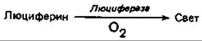
The specific substances involved in bioluminescence vary across different biological systems. This process is best understood in the American firefly Photinus pyralis, whose luciferin has been shown to be a benzothiazole derivative. Luciferase (E) catalyzes the reaction between reduced luciferin (LH2) and ATP; the resulting adenylate product emits light upon oxidation:

There is a direct linear correlation between the amount of ATP consumed in the reaction and the intensity of the emitted light. Consequently, the "firefly reaction" has become a widely favored method for the Quantitative determination of ATP.
Bacterial bioluminescence also involves multiple components: reduced FMN, O2, and a long-chain aldehyde (tetradecanal). The luciferase enzyme acts as a monooxygenase (see Section 14.11.4). The reaction can be expressed by the following equation:

The oxidation of FMNH2 presumably leads to the electronic excitation of FMN—specifically, the formation of an excited state [FMN ∙ H2O]—which emits light as it returns to the ground state:

Luminescence is widespread among Fungi (such as Armillaria mellea and Panus stipticus), Protozoa (dinoflagellates), and various Multicellular animals. In squids and certain deep-sea fish, light emission is mediated by symbiotic bacteria housed within specialized luminous Organs. While the ecological significance of bioluminescence in such animals is readily apparent, its biological function in unicellular organisms remains unclear.
Last update: 13/08/2026
Editorial and Educational Adaptation: This material has been compiled based on the primary/original source text. The project team performed an editorial review, corrected technical inaccuracies, structured sections, and adapted the content for an educational format.
What was processed:
- elimination of formatting defects (OCR errors, structural breaks, corrupted characters);
- editorial organization of content;
- standardization of terminology in accordance with academic sources;
- verification of factual statements against the original source text.
All mentions of the author, publication year, and origin of the primary text have been preserved in accordance with the source.