Tuberculosis Study Guide - M.M. Savula 2002
Secondary forms of pulmonary tuberculosis
Fibrocavitary pulmonary tuberculosis
Pathogenesis. This form of Pulmonary Tuberculosis is not the onset of the disease but rather the consequence of an unfavorable course of Other forms of tuberculosis: most commonly infiltrative or disseminated, less frequently Caseous Pneumonia, focal tuberculosis, or Tuberculoma, and very rarely, the Primary tuberculous complex. The cause of its development is late detection of tuberculosis or inadequate Treatment, which may be due to various factors: patient non-compliance, intolerance to anti-tuberculosis drugs, severe comorbidities, etc.
The Main Features of fibrocavitary pulmonary tuberculosis:
♦ an old fibrous cavity and scarring in the surrounding lung tissue;
♦ spread of infection through the Bronchi (bronchogenic dissemination);
♦ a prolonged course with alternating periods of exacerbation and remission;
♦ periodic or constant bacterial excretion, which makes this form always epidemiologically hazardous.
Morphological changes in fibrocavitary tuberculosis are highly characteristic (Fig. 19 - see Appendix). Its most important feature is an old fibrous cavity. Fibrosis is also pronounced around the cavity. It leads to deformation of the cavity and bronchi, development of Bronchiectasis, cirrhotic changes, and a reduction in the volume of the lobe or the entire lung. There may be a single cavity or several. As a result of the bronchogenic spread of mycobacteria, new foci are formed, usually below the cavity. They can coalesce to form infiltrates and break down to form new cavities ('daughter' cavities).
Clinical presentation. The Clinical symptoms of fibrocavitary tuberculosis depend on the extent of the process, its developmental phase, and complications. The medical history usually reveals that patients have been monitored and treated for tuberculosis for a long time. During exacerbations, symptoms become more pronounced: body Temperature rises, sweating (sometimes profuse night sweats) occurs, and appetite decreases. The most common complaint is a cough, ranging from a slight hack to an uncontrollable cough that disrupts the patient's Sleep. Sputum is difficult to expectorate; it is mucopurulent but lacks the foul odor characteristic of non-specific suppurative processes. Additionally, chest pain and shortness of breath may occur. Hemoptysis and even Pulmonary Hemorrhage occur periodically.
During remission, patients feel satisfactory and sometimes retain their ability to work. Over time, remissions shorten while exacerbations become more prolonged; patients lose weight, and Chronic Cor Pulmonale and cardiopulmonary insufficiency develop.
On physical examination, some patients do not differ from healthy individuals. In other cases, with a prolonged course of the disease, patients are pale and emaciated, showing retraction of the supraclavicular and infraclavicular fossae, narrowing of the affected side of the chest, and its lag during Respiration.
Over areas of scar tissue and infiltration zones, the Percussion note is dull. Auscultatory findings are sometimes minimal (in tuberculosis, 'much is seen' and 'little is heard'); in other cases, bronchial breathing and medium- or coarse-wet crackles are heard over areas of cirrhosis or a large cavity. Dry wheezes indicate concomitant Bronchitis.
Complete Blood count results differ between periods of remission and exacerbation. During exacerbations, leukocytosis (usually not exceeding 15-109/L), a left shift in the WHITE BLOOD Cell differential, lymphopenia, and a high ESR are observed. During remission, the blood count may be normal. The Mantoux test is positive. Pulmonary function is reduced, and chronic cor pulmonale gradually develops.
Mycobacterium tuberculosis is found in the sputum. If they are absent, especially in newly diagnosed cases, the Diagnosis of tuberculosis is not convincing.
Radiological changes are highly diverse. A characteristic feature is one or several cavities, at least one of which is inevitably an old, thick-walled, fibrous cavity, often deformed and surrounded by scar tissue bands, sometimes with cirrhotic changes in a volume-reduced lobe or the entire lung. The Mediastinum is sometimes shifted toward the affected side. Below the cavity, small focal shadows, infiltrates, and occasionally fresh 'daughter' destruction cavities are visible. If the initial form was disseminated tuberculosis, the process retains a certain Symmetry (Fig. 20).
Class="center">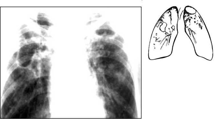
Fig. 20. Chest radiograph. Fibrocavitary pulmonary tuberculosis.
Fibrocavitary tuberculosis has a progressive course. It can lead to specific complications associated with the spread of tuberculous infection (Tuberculosis of the bronchus, Larynx, or intestines) and non-specific ones (chronic cor pulmonale, Amyloidosis of internal Organs, hemoptysis and hemorrhage, Spontaneous pneumothorax).
The Cytology/practical/136.html">Differential diagnosis OF fibrocavitary tuberculosis should be made with chronic abscess, lung cysts, and cavitating malignant tumor.
Treatment of patients with fibrocavitary pulmonary tuberculosis is complex. Combined antibacterial therapy is used, taking into account the sensitivity of the mycobacteria. Since there are irreversible destructive and cicatricial Changes in the Lungs, the effectiveness of therapy is low. It is possible to achieve a reduction in fresh inflammatory changes and destruction cavities, cessation of bacterial excretion, and improvement in the patient's general condition. In localized processes and with satisfactory functional parameters, Surgical treatment (resection) is used, followed by continued Chemotherapy.
Last update: 10/08/2026
Editorial and Educational Adaptation: This material has been compiled based on the primary/original source text. The project team performed an editorial review, corrected technical inaccuracies, structured sections, and adapted the content for an educational format.
What was processed:
- elimination of formatting defects (OCR errors, structural breaks, corrupted characters);
- editorial organization of content;
- standardization of terminology in accordance with academic sources;
- verification of factual statements against the original source text.
All mentions of the author, publication year, and origin of the primary text have been preserved in accordance with the source.