Human Physiology - William F. Ganong 2002
Blood Circulation
Nature of the Heartbeat and the Electrical Activity of the Heart
Origin and Spread of Electrical Impulses in the Heart
In the human Heart, the SA node is located near The entry of the SUPERIOR VENA CAVA into the right atrium. The AV node is situated in the right posterior region of the interatrial septum (Fig. 28-1). Three bundles of atrial fibers of the Purkinje fiber type emerge from the SA node and connect it to the AV node: the anterior internodal tract of Bachmann, the middle internodal tract of Wenckebach, and the posterior internodal tract of Thorel. However, debate continues regarding which specific structures mediate impulse transmission from the SA to the AV node: these named tracts or typical atrial cardiomyocytes. Under normal conditions, the AV node serves as the sole conducting pathway between the atria and ventricles. The AV node continues as the bundle of His. In the upper region of the interventricular septum, the bundle of His divides into right and left bundle branches. The left branch, in turn, gives rise to anterior and posterior fascicles. The branches and fascicles run subendocardially on both sides of the septum and transition into Purkinje fibers, which spread to all Regions of the ventricular myocardium.
The Histology of The Heart Muscle is described in Chapter 3. The conduction system is formed by atypical cardiomyocytes, which exhibit less distinct striations and fuzzy borders. The SA node and, to a lesser extent, the AV node also contain small, rounded Cells with few Organelles, interconnected by Gap Junctions. These are likely true pacemaker cells and are therefore referred to as P cells. The atrial muscle fibers are separated from the ventricular fibers by a ring of fibrous tissue. Normally, the only conducting pathway between the atria and ventricles is the bundle of His.
The SA node develops from structures on the right side of the embryo, whereas the AV node develops from structures on the left side. Consequently, in adults, the right Vagus nerve innervates primarily the SA node, while the left vagus nerve innervates the AV node. Similarly, right-sided sympathetic nerves supply the SA node, and left-sided sympathetic nerves supply the AV node. On both sides, most sympathetic fibers originate from the stellate ganglion. Noradrenergic fibers are epicardial, whereas vagal fibers are endocardial. However, interactions exist between the parasympathetic and sympathetic NERVES OF THE heart that produce reciprocal inhibitory effects. For instance, acetylcholine acts presynaptically to inhibit norepinephrine release by sympathetic nerve terminals, while neuropeptide Y, released by noradrenergic nerve endings, can suppress acetylcholine release.
Properties of the Heart Muscle
The electrical response of the heart muscle and nodal tissue, as well as the underlying ion movements, are described in detail in Chapter 3. Myocardial fibers have a Resting Potential of approximately -90 mV (see Fig. 3-15). The relationship between the Action Potential and contraction is illustrated in Fig. 3-14. Although the fibers are separated by membranes, gap junctions allow depolarization to spread radially across them as a syncytium. The transmembrane action potential of individual cardiomyocytes comprises phases of rapid depolarization, a plateau, and slow repolarization (see Fig. 3-15). Initial depolarization results from the influx of Na+ into The Cell through fast Na+ channels (Na+ current, INa). The influx of Ca2+ through slow Ca2+ channels (Ca2+ current, ICa) accounts for the plateau phase. Repolarization is driven by K+ efflux through Three types of K+ channels (see Chapter 3). The ECG represents the extracellularly recorded, summed electrical activity of all cardiac muscle fibers. The relationship between the action potentials of various regions of The Heart and the ECG tracing is shown in Fig. 28-1.
Class="center">
Fig. 28-1. Conduction system of the Heart. Typical transmembrane action potentials for the SA and AV nodes and other regions of the conduction system, as well as the atrial and ventricular Muscles, correlated with extracellularly recorded electrical activity, i.e., the Electrocardiogram (ECG). Action potentials and the ECG are plotted against a common time axis but have different baselines on the vertical axis; ALB = anterior left bundle branch.
Pacemaker Potentials
Following each impulse, the Membrane Potential of pacemaker cells reaches a threshold level. Consequently, this prepotential, or pacemaker potential (Fig. 28-2), triggers the subsequent impulse. At the peak of each impulse, IK is activated, causing repolarization. Subsequently, IK declines, and as K+ efflux from the cells decreases, membrane depolarization begins, driving the initial phase of the prepotential. Next, Ca2+ channels open. Two Types of these channels are distinguished in the heart: T-type (transient) channels and L-type (long-lasting) channels. The calcium current ICa through the opening of T-channels completes the prepotential, and ICa resulting from the opening of L-channels triggers the impulse.
The action potential in the SA and AV nodes is generated primarily via Ca2+ influx, with Na+ playing a less significant role. As a result, there is no phase of rapid depolarization characteristic of other regions of the conduction system, as well as the atrial and ventricular muscles (see Fig. 28-1). Under normal conditions, a prepotential is observed only in the SA and AV nodes. However, "latent pacemakers" exist in other PARTS OF THE conduction system that can manifest themselves if the SA and AV nodes are suppressed or if impulse transmission from them is impaired. Atrial and ventricular muscle fibers lack a prepotential and are capable of spontaneous depolarization only when injured or abnormal.
Upon stimulation of cholinergic vagal fibers supplying the nodes, the membrane hyperpolarizes, and the slope of the prepotential decreases. This is attributed to acetylcholine released by nerve terminals, which increases the K+ conductance of the nodal tissue. This effect is mediated by M2 muscarinic receptors, which, via G-protein β-γ subunits, induce the opening of K+ channels. Consequently, IKAch is generated, which counteracts the decline in IK. Furthermore, activation of M2 receptors reduces intracellular cAMP levels, leading to delayed opening of Ca2+ channels and, consequently, a reduction in impulse frequency. Strong vagal stimulation can temporarily halt spontaneous impulse generation.

Fig. 28-2. Membrane potential in pacemaker tissue. The current responsible for each phase of the potential is indicated below or nearby; L = long-lasting, T = transient. Note that the resting membrane potential of pacemaker tissue is slightly lower than that of atrial and ventricular muscles.
Conversely, stimulation of the cardiac sympathetic nerves promotes a more rapid decline in membrane potential, increasing the frequency of spontaneous impulse generation (Fig. 28-3). Norepinephrine released by sympathetic nerve endings binds to β1 receptors, elevating intracellular cAMP levels, opening L-channels, increasing ICa, and shortening the duration of the action potential depolarization phase.
In view of the innervation features described above, stimulation of the right vagus nerve slows heart rate primarily by inhibiting the SA node, whereas stimulation of the left vagus nerve mainly slows AV conduction. Similarly, stimulation of the right stellate ganglion accelerates heart rate, while stimulation of the left stellate ganglion accelerates AV conduction and decreases refractoriness.
The rate of impulse generation in the SA node and other regions of the conduction system can be modulated by Temperature and drugs. It increases with rising temperature, contributing to tachycardia in fever. Digitalis suppresses the conduction system and produces effects similar to vagal stimulation (particularly on the AV node).
Spread of Excitation in the Heart
Impulses originating in the SA node spread radially across the atria and converge on the AV node. Atrial depolarization is completed in approximately 0.1 s. Because impulse transmission through the AV node is slower (Table 28-1), There is a delay of about 0.1 s (AV nodal delay) before excitation spreads to the ventricles. This delay is shortened by sympathetic nerve stimulation and prolonged by vagal stimulation. From the apex of the septum, the wave of depolarization rapidly propagates via Purkinje fibers to all regions of the ventricles (within 0.08–0.1 s). In humans, ventricular muscle depolarization begins on the left side of the interventricular septum and initially spreads rightward across the mid-septum. The depolarization wave then travels down the septum toward the apex of the heart and turns back along the ventricular walls toward the AV groove, spreading from the endocardial surface to the epicardial surface (Fig. 28-4). Impulses reach the posterior wall of the left ventricle, the Base of the heart, the pulmonary conus, and the upper region of the septum last.
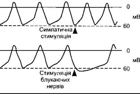
Fig. 28-3. Effect of sympathetic (noradrenergic) and vagal (cholinergic) nerve stimulation on the membrane potential in the SA node.
Last update: 10/08/2026
Editorial and Educational Adaptation: This material has been compiled based on the primary/original source text. The project team performed an editorial review, corrected technical inaccuracies, structured sections, and adapted the content for an educational format.
What was processed:
- elimination of formatting defects (OCR errors, structural breaks, corrupted characters);
- editorial organization of content;
- standardization of terminology in accordance with academic sources;
- verification of factual statements against the original source text.
All mentions of the author, publication year, and origin of the primary text have been preserved in accordance with the source.