Review of Medical Physiology - William F. Ganong 2002
Physiology of Nerve and Muscle Cells
Excitatory Tissue: Nerve
Ionic Basis of Excitation and Conduction
Neuronal Cell membranes, much like those of other Cells, feature A wide variety of Ion Channels. Some of these are passive, meaning they remain constantly open, whereas others are voltage-gated or Ligand-gated. The function of these channels—specifically Na+ and K+ channels—accounts for the electrical phenomena observed in nerves.
Ionic Basis of the Resting Membrane Potential
As noted in Chapter 1, Neurons and other cells actively transport Na+ outward while taking in K+. Through diffusion, K+ leaks out of cells and Na+ diffuses inward; however, thanks to K+ channels, membrane permeability is significantly higher for K+ than for Na+. Because the membrane is impermeable to most intracellular anions, the efflux of K+ is not accompanied by a corresponding outflow of anions, thereby generating membrane polarization. Its outer surface carries a positive charge, while the inner surface holds a negative charge.
Ionic Fluxes During the Action Potential
Changes in membrane permeability to Na+ and K+ occurring during an action potential are illustrated in Fig. 2-12. The permeability for an ion is inversely proportional to its electrical resistance within the membrane and serves as a measure of that ion's penetrability.
A slight decrease in the resting membrane potential prompts an outward flow of K+ and an influx of Cl-, which serves to restore the resting membrane potential. However, when depolarization reaches 7 mV, the conductance of voltage-gated Na+ channels increases (Na+ channel activation). Once this critical depolarization threshold is reached, membrane permeability to Na+ increases so dramatically that Na+ rushes into the fiber in an avalanche-like manner, Setting up high concentration and electrical gradients that temporarily override the repolarizing factors. The equilibrium potential for Na+ in mammalian neurons, calculated using the Nernst equation, is approximately +60 mV. The membrane potential approaches this value, though it is not fully reached during the action potential, primarily because the increase in Na+ permeability is short-lived. The gates of the Na+ channels rapidly close (inactivation state), lasting a few milliseconds until the resting membrane potential is recovered. Furthermore, the direction of the electrical gradient for Na+ reverses during the overshoot due to the changing membrane potential, which limits further Na+ entry into the axon. A third factor driving repolarization is the activation and opening of K+ channel gates, which takes longer than Na+ channel activation; consequently, the peak of K+ conductance occurs later than that of Na+. The net outward movement of positive charges driven by K+ efflux completes the repolarization process. The phenomenon of after-hyperpolarization is attributed to the slow inactivation of K+ channels.
Class="center">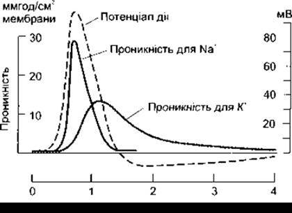
Fig. 2-12. Changes in membrane permeability to Na+ and K+ during The formation of an action potential in a giant squid axon. The dashed line tracing the action potential curve is plotted against the time scale. As shown, the initial electrotonic depolarization triggers A change in Na+ permeability, which in turn promotes depolarization (reproduced by permission from Hodkin AL. Ionic movements and electrical activity in giant nerve fibers. Proc R Soc Long Ser B 1958; 143.1).
A decrease in extracellular Na+ concentration leads to a smaller action potential, yet it has little effect on the resting membrane potential. This minor influence on the Resting Potential can be explained by the Goldman Equation (see Chapter 1), given that resting membrane permeability to Na+ is relatively low. Conversely, an elevation in extracellular K+ concentration decreases the resting membrane potential.
Although Na+ enters Nerve Cells and K+ leaves them during an action potential, the actual number of ions involved in these movements is negligible compared to their total intracellular pool.
The fact that a nerve takes up Na+ and loses K+ during activity has been confirmed experimentally, though a noticeable difference is detected only after prolonged, repetitive stimulation.
The slow opening and closing kinetics of K+ channel gates also account for the phenomenon of axonal accommodation. If depolarization occurs rapidly, the opening of Na+ channels overwhelms the repolarizing forces; however, if the induced depolarization is slow, the opening of K+ channels has enough time to counterbalance the gradual opening of Na+ channels, preventing an action potential from firing.
A decrease in extracellular Ca2+ concentration enhances the excitability of nerve and Muscle cells by lowering the threshold of depolarization required to trigger changes in Na+ and K+ permeability and elicit an action potential. Conversely, an elevation in extracellular Ca2+ stabilizes the membrane by reducing excitability.
Distribution of Ion Channels in Myelinated Neurons
As mentioned in Chapter 1, specific substances that bind to Na+ and K+ channels can be tagged, labeled, and used to identify the localization of these channels within The Cell membrane. Voltage-gated Na+ channels are heavily concentrated at the nodes of Ranvier and the initial segments of myelinated neurons. The initial segment and the first node of Ranvier in sensory neurons serve as the sites where impulses are generated. Other nodes of Ranvier act as the loci where saltatory conduction takes place. The density of Na+ channels per 1 µm2 of a mammalian myelinated neuron is 50–75 on the cell body, 350–500 at the initial segment, up to 25 along the myelin surface, 2,000–12,000 at the node of Ranvier, and 20–25 at the axon terminals. On unmyelinated axons, this density is approximately 110. In many myelinated neurons, Na+ channels lie adjacent to K+ channels, which play a crucial role in repolarization.
Energy Sources and Nerve METABOLISM
The majority of a nerve's Energy Requirements—approximately 70%—are dedicated to maintaining the polarized state of the membrane via The activity of Na+-K+-ATPase. During maximal activity, the metabolic rate of a nerve doubles (by comparison, the metabolic rate of Skeletal Muscle increases a hundredfold). Inhibition of lactic acid production does not affect Muscle Function.
Much like muscle, a nerve generates heat at rest, initial heat during the action potential, and delayed heat following activity. However, in a nerve, the delayed heat produced after a single impulse is 30 times greater than the initial heat. Evidence suggests that initial heat output is higher during after-depolarization than during the spike of the action potential. Muscle metabolism is covered in detail in Chapter 3.
Last update: 10/08/2026
Editorial and Educational Adaptation: This material has been compiled based on the primary/original source text. The project team performed an editorial review, corrected technical inaccuracies, structured sections, and adapted the content for an educational format.
What was processed:
- elimination of formatting defects (OCR errors, structural breaks, corrupted characters);
- editorial organization of content;
- standardization of terminology in accordance with academic sources;
- verification of factual statements against the original source text.
All mentions of the author, publication year, and origin of the primary text have been preserved in accordance with the source.