Medical Radiology - Lazar A.P. 2008
Radiation Examination of the Genitourinary System
Radiation Diagnostics of Diseases of the Internal Female Genital Organs
A normal Uterus is pear-shaped and located posterior to the Urinary Bladder. The dimensions of its body depend on the number of previous pregnancies and deliveries, averaging 6-8 cm in length, 3.5-4.5 cm in thickness, and 4.5-6.5 cm in width. The length of the cervix is 2-3.5 cm, and the cervicouterine index is 1/2. The Ovaries lie adjacent to the lateral walls of the uterus, measuring 2-3.5 cm, with an oval shape and distinct margins.
Ultrasound examination of the Internal female reproductive Organs has no contraindications and can be performed during any phase of the Menstrual cycle or at any stage of Pregnancy. Prior to sonography, the urinary bladder must be filled; this displaces the intestinal loops out of the pelvic cavity and acts as an acoustic window for the propagation of ultrasound waves. The Location OF THE uterus and ovaries (Fig. 256), their shape, endometrial state, and echogenicity are determined, linear dimensions are measured, margins and Structure are evaluated, and other pelvic organs are visualized, followed by the characterization of any Structural Features of detected pathological formations (cysts, tumors, Ectopic Pregnancy, etc.) (Fig. 257).
Class="center">
Fig. 257. Ectopic pregnancy on transvaginal ultrasound.
1 - yolk sac of the embryo located within a thick-walled fallopian tube (3);
2 - uterus.
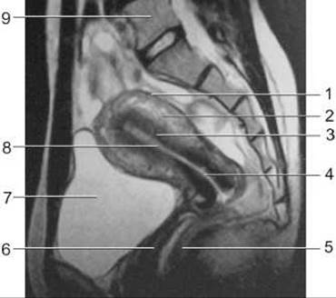
Fig. 258. Magnetic Resonance imaging (MRI) of the female pelvic organs in the sagittal plane.
1 - perimetrium;
2 - myometrium;
3 - endometrium;
4 - cervical canal;
5 - rectum;
6 - Vagina;
7 - urinary bladder;
8 - uterine cavity;
9 - 5th lumbar vertebra.
Computed tomography (CT) of the female pelvis is performed after ultrasound to determine THE ORIGIN OF a space-occupying lesion, assess its extent and relationship with surrounding organs, and evaluate Lymph node enlargement. The CT scan is performed after bowel cleansing. If it is necessary to differentiate the intestine from pathological masses, especially large ones, contrast enhancement is used: 3 to 4 hours before the CT scan, the patient is asked to drink 200 ml of a 0.5% Urografin solution to fill the terminal ileum; immediately before the Procedure, 100 ml of Urografin is administered rectally; and 20 to 30 minutes prior to the scan, the patient drinks 2 glasses of Water to fill the urinary bladder.
On computed tomography, a homogeneously structured uterus with a density of +40 to +60 HU is visualized in the center of the pelvis. The ovaries are located posterior to the round ligaments extending from the uterine horns to the pelvic walls.
CT image enhancement via intravenous administration of radiocontrast agents increases the densitometric difference between healthy and pathologically altered tissue due to differences in their Blood supply.
Magnetic resonance imaging of the FEMALE REPRODUCTIVE ORGANS is performed without preparation, with the patient in a supine position during shallow breathing. On T1-weighted images, the uterus has a homogeneous structure, clear margins, and produces a medium-intensity signal. On T2-weighted images, the internal architecture of the uterus can be differentiated: the endometrium produces a high-intensity signal, the myometrium yields a medium-intensity signal in the thick outer zone and a low-intensity signal in the narrow inner zone, and the cervix also produces a low-intensity signal due to the predominance of Connective Tissue (similar to the inner zone of the myometrium); the cervical canal, filled with mucus, produces a high-intensity signal (Fig. 258). Ovaries are identified more frequently on MRI than on CT. On T1-weighted images, they produce a low-intensity signal. On T2-weighted images, the ovarian stroma produces a low-intensity signal, whereas follicles produce a high-intensity signal.

Fig. 259. Hysterosalpingography (B, C - diagram).
A, B - anteroposterior projection; C - left lateral projection;
1 - uterine cannula; 2 - isthmus; 3 - uterine cavity; 4 - uterine horn; 5 - uterine fundus; 6 - uterotubal junction; 7 - fallopian tube; 8 - ampulla of the fallopian tube; 9 - contrast medium in the peritoneal cavity.
Radiological examination of the Internal Female Genitalia is performed using contrast media and is referred to as metrosalpingography or hysterosalpingography. Indications for hysterosalpingography include suspected tubal occlusion in Infertility, malformations, Tuberculosis of the uterus and appendages, polyps, submucous myomas, and Endometriosis. Hysterosalpingography is contraindicated in acute or chronic local infections, Trichomoniasis, severe general condition, pregnancy, and vaginal cleanliness grades III and IV. It is best performed between the 10th and 20th days of the menstrual cycle to prevent the contrast agent from entering gaping Blood Vessels and introducing infection.
To perform hysterosalpingography, a uterine nozzle or catheter is inserted into the uterine cavity through the cervical canal. Using a syringe, about 10 mL of a water-soluble radiopaque agent (such as urographin, cardiotrast, omnipaque, etc.) is slowly injected through the catheter. Oil-based contrast media, which were used previously, pass slowly through the fallopian tubes and can cause aseptic tubal inflammation with subsequent occlusion; therefore, they are no longer in use. Radiographs are taken in the anteroposterior projection immediately after contrast administration, and again at 10 and 30 minutes. Normally, on the final radiograph, the contrast medium should appear in the Abdominal cavity as scattered droplets. The healthy uterine cavity appears as a symmetrical triangle with its base directed upward and elongated horns (Fig. 259).

Fig. 260. Sonography at 7 weeks of gestation (diagram).
1 - pregnant uterus;
2 - gestational sac surrounded by an echogenic layer of trophoblast;
3 - embryo, whose length allows Determination of Gestational Age;
4 - filled urinary bladder;
5 - rectum;
6 - sacrum.
Radionuclide metrosalpingography is performed by injecting 1 mL of radiopharmaceutical into the uterine cavity. A clamp is applied to the cervix, and scintigraphy is performed 30 minutes later. On a normal scintigram, the radiopharmaceutical is evenly distributed within the uterine cavity and appendages. The indication for radionuclide metrosalpingography, similar to X-ray hysterosalpingography, is primarily the evaluation of fallopian tube patency.
Radioimmunoassay allows for the detection of blood concentrations of Hormones produced by the female reproductive organs (estradiol, progesterone), Pituitary Hormones regulating female reproductive function (prolactin, lutropin, follitropin), hypothalamic releasing hormones regulating pituitary hormone production (luliberin, foliberin), as well as the identification of complex hormonal imbalances that affect the function of the female genital organs.
Pregnancy can be sonographically detected as early as 5-6 weeks based on the visualization of an asynchronously positioned gestational sac in the uterus, featuring an anechoic central part and an echogenic periphery. At 6 weeks, the gestational sac occupies 1/3 of the uterine cavity volume, and by 8-9 weeks, it occupies nearly half of the cavity.
At 7-8 weeks, an embryo measuring 9-10 mm can be visualized (Fig. 260), and by 9-10 weeks, its HEAD and body become discernible. Fetal sex can be determined between 24 and 34 weeks of gestation by identifying the Penis and Scrotum in male fetuses. In later stages, their detection becomes difficult due to a decrease in Amniotic Fluid volume and the large size of the fetus.
In obstetrics, determining the state of the Placenta and its relation to the internal os of the cervix is of vital importance. Most commonly, it is located on the anterior or posterior wall of the uterus, extending onto the lateral wall. The placenta begins to be visualized in early second trimester as a flat, echogenic structure with a granular texture, while placental cotyledons become noticeable in the third trimester. If the distance from the lower edge of the placenta to the internal cervical os is less than 7 cm, low-lying placenta or Placenta Previa is diagnosed, which poses a danger to the life of both mother and fetus. In premature placental abruption, a retroplacental hematoma appears between the placenta and the uterine wall as an anechoic structure. Sonographic signs of fetal demise include the absence of fetal movements and Heart activity, lag in uterine size relative to gestational age, and deformation of the gestational sac.

Fig. 261. Endometriosis on ultrasound using a transvaginal probe. The arrow points to the Ovary with follicles. Endometriosis (1) and a cyst (2) adjacent to the ovary.
Endometriosis is defined as the presence of secretory endometrium in an ectopic location. It involves glandular-cystic hyperplasia of the uterine mucosa, typically developing after uterine trauma (surgical interventions, cervical diathermy coagulation, etc.). Endometriosis can affect organs adjacent to the uterus, most commonly the ovaries. Using ultrasound, CT, and MRI, uterine enlargement and the presence of endometriomas can be detected. On echograms, endometriomas appear as cysts with diffuse low-amplitude internal echoes, a prominent wall, and enhanced acoustic transmission (Fig. 261). During ultrasound, small 2-5 mm round anechoic structures that enlarge prior to the menstrual cycle may also be detected in the myometrium. On MRI, affected areas produce a high-intensity signal. Endometriosis is best diagnosed via laparoscopy.
Uterine tumors can be benign or malignant. Among benign tumors, fibroids (leiomyomas) are the most common, resulting from hypertrophy and proliferation of connective and muscular tissue elements. On ultrasound, uterine fibroids manifest as uterine enlargement, deformation, and additional masses, typically of reduced echogenicity. On hysterosalpingography, a fibroid deforms the uterine cavity, showing as a round filling defect with sharp, smooth contours, unlike Uterine body Cancer, which presents with uneven, indistinct contours (Fig. 262). Magnetic resonance imaging allows for a better Assessment of the number, size, and depth of the fibroid than ultrasound, CT, or hysterosalpingography (Fig. 263). The intensity of the MR signal depends on the predominance of fibrous or muscular tissue within the tumor structure, as well as the presence of necrosis and cystic cavities.

Fig. 262. Uterine tumors on hysterosalpingogram (diagram).
A - fibroid as a round filling defect with smooth contours;
B - Cancer of the uterine fundus as a filling defect with irregular, jagged, indistinct contours.
Carcinoma of the uterine body is sonographically detected most frequently as an echonegative node, which may be heterogeneous due to necrosis and Hemorrhage. Ultrasound can identify cancer nodes larger than 1 cm, while smaller nodes can be detected using MRI. CT and MRI not only identify the cancerous tumor but also reveal its invasion into adjacent structures and metastases in pelvic Lymph Nodes. Artificial contrast enhancement AIDS in detection. CT is also crucial for planning and evaluating the efficacy of Radiation therapy.
Ovarian cancer on echograms and computed tomograms is identified by the irregular shape of the tumor, indistinct contours, heterogeneous internal structure, and infiltration of the soft pelvic Tissues. Ultrasound- or CT-guided needle biopsy of the ovary aids in Diagnosis.
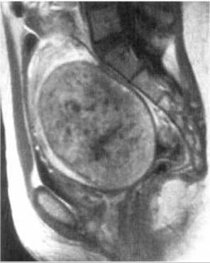
Fig. 263. Uterine Fibroid on MRI (T1-weighted image).
On ultrasound, Ovarian Cysts appear as echonegative structures with a thin capsule, clear smooth margins, and posterior acoustic enhancement. Computed tomography reveals a round cyst with a thin, smooth wall, distinct margins, low attenuation (+5 to +12 HU), and no soft tissue infiltration or enlarged regional lymph nodes.
Last update: 08/08/2026
Editorial and Educational Adaptation: This material has been compiled based on the primary/original source text. The project team performed an editorial review, corrected technical inaccuracies, structured sections, and adapted the content for an educational format.
What was processed:
- elimination of formatting defects (OCR errors, structural breaks, corrupted characters);
- editorial organization of content;
- standardization of terminology in accordance with academic sources;
- verification of factual statements against the original source text.
All mentions of the author, publication year, and origin of the primary text have been preserved in accordance with the source.