Human Anatomy - Kotsan I. Ya. 2009
Respiratory System
External Nose and Nasal Cavity
The Nose (nasus) is the initial segment of the Respiratory system. It is divided into the External Nose and the Nasal cavity (Fig. 162).
The external nose (nasus externus) consists of bony and cartilaginous structures covered by Skin. In shape, it resembles a triangular pyramid, one face of which (the posterior) is absent because the posterior edges of the lateral faces merge directly into the facial surface. The shape of the external nose exhibits a very high degree of Variability determined by heredity. The external nose comprises the ROOT of the nose (radix nasi), located superiorly; the apex of the nose (apex nasi), directed inferiorly; and two lateral sides that converge along the midline to form the dorsum of the nose (dorsum nasi), facing anteriorly. The lower PARTS OF THE lateral sides of the nose are called the alae of the nose (alae nasi). The lower edges of the alae bound the nasal apertures — the nostrils (nares) — which serve for the passage of air into and out of the nasal cavity. Unlike the nostrils of animals, including primates, human nostrils face inferiorly rather than anteriorly. As a result, the stream of inhaled air is directed not straight backward like in apes, but upward toward the olfactory region, following a long, arch-like path to the nasopharynx, which facilitates the conditioning of the air (moisturization, purification, and warming). Exhaled air, in contrast, passes in a straight line through the inferior nasal meatus. The protruding external nose is a specific human feature associated with the upright posture of the body and the transformations of the facial Skeleton caused, on the one hand, by the reduction of masticatory function and, on the other hand, by The Development of speech.
Class="center">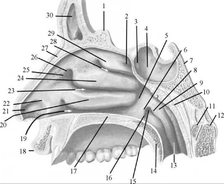
Fig. 162. Nasal cavity (lateral wall)
1 — crista galli; 2 — sphenoethmoidal recess; 3 — aperture of sphenoidal sinus; 4 — sphenoidal sinus; 5 — nasopharyngeal meatus; 6 — anterior lip; 7 — clivus of Occipital bone; 8 — posterior lip; 9 — tubal elevation; 10 — pharyngeal tonsil; 11 — anterior arch of atlas; 12 — axis (vertebra); 13 — salpingopharyngeal fold; 14 — soft palate; 15 — pharyngeal opening of auditory tube; 16 — salpingopalatine fold; 17 — hard palate; 18 — upper lip; 19 — inferior nasal meatus; 20 — Inferior nasal concha; 21 — nasal vestibule; 22 — limen nasi; 23 — middle nasal meatus; 24 — middle nasal concha; 25 — vestibule of middle meatus; 26 — agger nasi; 27 — Nasal bone; 28 — superior nasal meatus; 29 — superior nasal concha; 30 — frontal sinus
The bony Skeleton of the external nose is formed by the nasal bones and the frontal processes of the maxillae. The cartilaginous skeleton of the nose consists of the nasal cartilages (cartilagines nasi), which are composed of hyaline Cartilage and are interconnected and joined to the bones by Connective Tissue. The following nasal cartilages are distinguished.
The lateral nasal cartilage (cartilago nasi lateralis) is a paired, triangular cartilage located directly beneath the nasal bone, participating in The formation of the lateral wall of the external nose. The anterior edges of the right and left lateral nasal cartilages join along the midline to form the dorsum of the nose.
The major alar cartilage (cartilago alaris major) is a paired cartilage situated inferior to the lateral nasal cartilage, bounding the nostril anteriorly and laterally.
The minor alar cartilages (cartilagines alares minores) are small cartilages, numbering two to three on each side, located posterior to the major alar cartilage, between it and the margin of the piriform aperture.
The septal nasal cartilage (cartilago septi nasi) is an unpaired, irregularly quadrangular cartilage positioned vertically, forming part of the anterior nasal septum. Posterosuperiorly, it connects with the perpendicular plate of the Ethmoid bone, and posteroinferiorly, with the Vomer and the anterior nasal spine.
The vomeronasal cartilage (cartilago vomeronasalis) is a paired, narrow strip of cartilage situated between the inferior margin of the septal nasal cartilage and the anterior margin of the vomer.
Occasionally, between the major alar cartilage and the septal nasal cartilage, there are several inconstant, variable-sized accessory nasal cartilages (cartilagines nasales accessoriae).
The nasal cavity (cavitas nasi) along with its air-filled Paranasal Sinuses (sinus paranasales)—the maxillary, sphenoid, frontal, and ethmoid air Cells—serves as the site where inhaled air is warmed, moisturized, and filtered of dust before reaching the Lungs. Anteriorly, the nasal cavity opens to the external environment, and posteriorly, it communicates with the nasopharynx. It communicates with the outside via the nasal apertures (nostrils) and with the nasopharynx via the choanae.
The nasal cavity features inferior, anterosuperior, and lateral walls. The inferior wall is formed by the palatine process of the Maxilla and the horizontal plate of the Palatine bone; the anterosuperior wall by the cribriform plate of the ethmoid bone, as well as the nasal and frontal bones; and the lateral walls by the maxilla, lacrimal and ethmoid bones, inferior nasal concha, perpendicular plate of the palatine bone, and the Medial surface of the pterygoid process of the Sphenoid bone. The nasal septum (septum nasi) divides the nasal cavity into right and left halves. The nasal septum is formed by the perpendicular plate of the ethmoid bone, the vomer, and is completed anteriorly by cartilage.
On the lateral walls of the nasal cavity, there are three nasal conchae: superior, middle, and inferior (concha nasalis superior, concha nasalis media, and concha nasalis inferior). The first two conchae are outgrowths of the ethmoid bone, whereas the inferior one is a separate bone. The presence of the nasal conchae increases the internal surface area of the nasal cavity that comes into contact with the air. The spaces between the conchae are called nasal meatuses: superior (meatus nasi superior), middle (meatus nasi medius), and inferior (meatus nasi inferior). The apertures of the paranasal sinuses open into the nasal meatuses. The sphenoid sinus and posterior ethmoid air cells open into the superior nasal meatus; the middle and anterior ethmoid air cells, frontal and maxillary sinuses open into the middle nasal meatus; and the nasolacrimal canal, through which the lacrimal sac communicates with the nasal cavity, opens into the inferior nasal meatus. The length of the nasolacrimal canal is 12–24 mm.
Within the nasal cavity, a distinction is made between the nasal vestibule (vestibulum nasi) and the nasal cavity proper (cavum nasi proprium). The nasal vestibule is separated from the nasal cavity proper by a slight ridge, the limen nasi, formed by the upper edge of the lateral crus of the major alar cartilage. The internal surface of the nasal vestibule is lined with skin continuous with that of the external nose through the nostrils, and it contains hairs, Sebaceous Glands, and Sweat Glands.
The nasal cavity proper is lined with a mucous membrane, which is divided into two structurally and functionally distinct regions: the respiratory and olfactory regions.
The respiratory region (regio respiratoria) is covered by ciliated Cytology/practical/33.html">Pseudostratified columnar epithelium, whose cilia form a continuous carpet where dust particles settle. The beating of the cilia moves the trapped dust out of the nasal cavity. Additionally, the mucous membrane contains mucous glands whose secretion envelops dust and AIDS in its removal, as well as humidifying the inhaled air. In the region of the middle and inferior nasal conchae, the mucosa and submucosa contain A large number of venous vessels that form cavernous venous plexuses of the conchae (plexus (venosi) cavernosi concharum), which help to warm the incoming air.
The olfactory region (regio olfactoria) of the nasal mucosa contains the peripheral nerve endings of the Olfactory nerve—specialized olfactory neurosecretory bipolar cells that act as the receptors of the olfactory analyzer. That part of the mucous membrane lining the superior nasal conchae, the free surfaces of the middle conchae facing the nasal septum, and the corresponding superior section of the nasal septum belongs to the olfactory region. All the remaining portions of the mucous membrane are part of the respiratory region. When the nostrils are destroyed by some pathological process and the airflow during respiration bypasses the inferior nasal meatus—that is, fails to come into contact with the olfactory region—a person cannot perceive odors.
Blood supply to the external nose and the walls of the nasal cavity is provided by Branches of the maxillary and ophthalmic Arteries. Venous blood drains into the facial vein and the pterygoid venous plexus. Lymphatic vessels from the mucous membrane of the nasal cavity lead to the submandibular and submental Lymph Nodes.
Sensory Innervation of the mucous membrane of the nose (its anterior part) is supplied by branches of the anterior ethmoidal nerve from the nasociliary nerve. The posterior part of the lateral wall is innervated by branches of the nasopalatine nerve and posterior nasal branches from the maxillary nerve. The Glands of the nasal mucosa are innervated from the pterygopalatine ganglion via posterior nasal branches and the nasopalatine nerve, originating from the autonomic Nucleus of the intermediate nerve from the Facial Nerve.
Age-related Features of the nasal cavity. The nose of a newborn infant is flattened and short, and the nasal cavity is narrow and low. With age, the dorsum of the nose lengthens, and the tip of the nose is formed. During Puberty, the shape of the nose becomes permanent.
The nasal conchae in newborns are relatively thick. The superior nasal meatus is absent, while the middle and inferior ones are poorly developed. The middle nasal meatus forms by six months of age, the inferior by two years, and the superior after two years of age.
Of the paranasal sinuses, only the maxillary sinus is present at birth. The remaining sinuses begin to develop after birth. The frontal sinus appears In the second year of life, the sphenoid sinus in the third year, and the ethmoid air cells between three and six years of age.
Last update: 08/08/2026
Editorial and Educational Adaptation: This material has been compiled based on the primary/original source text. The project team performed an editorial review, corrected technical inaccuracies, structured sections, and adapted the content for an educational format.
What was processed:
- elimination of formatting defects (OCR errors, structural breaks, corrupted characters);
- editorial organization of content;
- standardization of terminology in accordance with academic sources;
- verification of factual statements against the original source text.
All mentions of the author, publication year, and origin of the primary text have been preserved in accordance with the source.