Human Anatomy - Kotsan I. Ya. 2009
Respiratory System
Larynx
Air from the Nasal cavity passes through the choanae into the nasal part of the Pharynx, then into the oral part of the pharynx, and subsequently into the Larynx. In addition to conducting air, the larynx is involved in phonation, articulate speech production, and the act of swallowing.
The larynx is located in the anterior region of the neck at the level of the IV—VI cervical vertebrae. Superiorly, it is attached to the Hyoid bone via the thyrohyoid membrane (membrana thyrohyoidea), and inferiorly it connects with the Trachea. Anteriorly, the larynx is covered by the Infrahyoid Muscles; laterally, it is flanked by major Blood Vessels and the lobes of The Thyroid Gland; posteriorly, it is adjacent to the pharynx. Air enters the larynx from the pharynx through the laryngeal inlet (aditus laryngis), which is bounded anteriorly by the epiglottis, posteriorly by the apices of the arytenoid cartilages, and laterally by the aryepiglottic folds (plicae aryepiglotticae), connecting the right and left arytenoid cartilages to the respective margins of the epiglottis.
The laryngeal cavity (cavitas laryngis) consists of three parts: the upper—the laryngeal vestibule, the middle—the intermediate cavity of the larynx, and the lower—the infraglottic cavity (Fig. 163).
Class="center">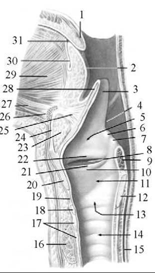
Fig. 163. Laryngeal cavity, right side (according to R. D. Sinelnikov)
1 — uvula (palatine); 2 — ROOT of the Tongue; 3 — epiglottis; 4 — aryepiglottic fold; 5 — laryngeal vestibule; 6 — cuneiform tubercle; 7 — corniculate tubercle; 8 — laryngeal ventricle; 9 — arytenoid muscles (transected); 10 — vocal fold; 11 — infraglottic cavity; 12 — lamina of the cricoid Cartilage; 13 — laryngeal cavity; 14 — trachea; 15 — Esophagus; 16 — thyroid gland; 17 — tracheal cartilages; 18 — arch of the cricoid cartilage; 19 — cricothyroid ligament; 20 — thyroid cartilage; 21 — vocal fold; 22 — vestibular fold; 23 — adipose tissue; 24 — median thyrohyoid ligament; 25 — hyoepiglottic ligament; 26 — hyoid bone (body); 27 — geniohyoid Muscle; 28 — epiglottic cartilage; 29 — genioglossus muscle; 30 — remnant of the thyroglossal duct; 31 — cecal foramen of the tongue
The laryngeal vestibule (vestibulum laryngis) extends from the laryngeal inlet to the vestibular folds (plicae vestibulares), between which lies the rima vestibuli. The anterior wall of the vestibule is formed by the mucous membrane-covered epiglottis, and the posterior wall by the arytenoid cartilages. The height of the anterior wall of the vestibule reaches 4 cm, while the posterior wall is 1.0—1.5 cm high.
The middle section—the intermediate cavity of the larynx (cavitas laryngis intermedia)—has the most complex Structure, extending from the vestibular folds above to the vocal folds (plicae vocales) below. Between the vestibular fold and the vocal fold on each side of the larynx lies a small depression—the laryngeal ventricle (ventriculus laryngis), which acts as a resonator during sound production. The rima glottidis is located between the right and left vocal folds. The larger anterior part of the rima glottidis, corresponding to THE POSITION OF the right and left vocal cords, is called the intermembranous part (pars intermembranacea), whereas the smaller posterior part, situated between the arytenoid cartilages, is termed the intercartilaginous part (pars intercartilaginous). The length of the rima glottidis (anteroposterior dimension) is 20—24 mm in men and 16—19 mm in women, with the membranous part accounting for 15 mm in men and 12 mm in women. The width of the rima glottidis during quiet breathing is 5 mm. Sound volume and pitch depend on the width of the rima glottidis: a widened rima glottidis produces low-pitched sounds, while a narrowed one produces high-pitched sounds.
The lower division of the laryngeal cavity—the infraglottic cavity (cavitas infraglottica)—is situated below the rima glottidis; inferiorly, this section widens in a cone-like fashion and passes directly into the tracheal cavity.
The wall of the larynx is formed by mucous, fibrocartilaginous, and adventitial membranes.
The mucous membrane lines the internal surface of the larynx. Most of it is covered by ciliated epithelium, except for the posterior surface of the epiglottis, the region of the vocal folds, and the inner surface of the arytenoid cartilages, which feature stratified squamous epithelium. The mucous membrane is pink in color and contains seromucous glands, which are particularly abundant in the area of the vestibular folds and laryngeal ventricles. The secretion of these glands moistens the vocal folds, traps dust, and the cilia of the ciliated epithelium expel it outward through coughing movements.
The submucosa of the larynx contains A large number of fibrous and elastic fibers that form the fibroelastic membrane of the larynx (membrana fibroelastica laryngis). This membrane comprises two parts: the quadrangular membrane (membrana quadrangularis), located in the anterosuperior region of the larynx, and the elastic cone (conus elasticus), situated in the lower level of the larynx. The quadrangular membrane participates in forming the wall of the vestibule. Superiorly, it reaches the aryepiglottic folds, and its lower edge contributes to The formation of the vestibular ligaments (ligg. vestibulares), located within the folds of the same name. The elastic cone is a continuation of the cricothyroid ligament; its upper free edge helps form the vocal ligaments (ligg. vocales).
The fibrocartilaginous membrane consists of hyaline and elastic cartilages surrounded by dense Fibrous Connective Tissue and muscles, serving as the framework (Skeleton) of the larynx (Fig. 164). The laryngeal cartilages include three unpaired (thyroid, cricoid, epiglottic) and three paired (arytenoid, corniculate, cuneiform) cartilages. The epiglottic, corniculate, and cuneiform cartilages are composed of elastic cartilage, whereas the thyroid, cricoid, and most of the arytenoid cartilages consist of hyaline cartilage.
The thyroid cartilage (cartilago thyroidea) is the largest of the laryngeal cartilages. It contributes to the Formation of the anterior and lateral walls of the larynx. The thyroid cartilage consists of two quadrangular plates: the right and left (lamina dextra et lamina sinistra), which fuse anteriorly at an angle of 120° in women and 90° in men. The laryngeal prominence formed by this angle is more pronounced in men (the "Adam's apple," a secondary male sex characteristic). The upper edge of the thyroid cartilage angle projects further anteriorly than the lower edge and features the superior thyroid notch (incisura thyroidea superior), to which the lower end of the epiglottis is attached by a ligament. On the lower border of the thyroid cartilage, at the junction of the two plates, There is a weakly defined inferior thyroid notch (incisura thyroidea inferior). The external surface of both plates bears an oblique line (linea obliqua)—the attachment site for the sternothyroid and thyrohyoid muscles. The oblique line terminates superiorly at the superior thyroid tubercle (tuberculum thyroideum superius) and inferiorly at the inferior thyroid tubercle (tuberculum thyroideum inferius). The posterior free margin of each plate is thickened and features upward- and downward-directed processes termed the superior and inferior horns (cornu superius et cornu inferius), respectively. The superior horns connect the thyroid cartilage to the hyoid bone via ligaments, while the inferior horns articulate via joints with the cricoid cartilage.
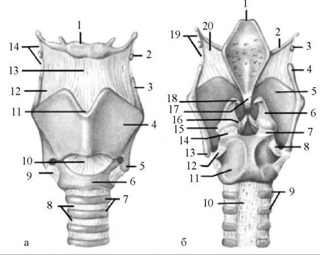
Fig. 164. Cartilages, ligaments, and JOINTS OF THE larynx
a — anterior view: 1 — hyoid bone; 2 — triticeal cartilage; 3 — superior horn of the thyroid cartilage; 4 — left lamina of the thyroid cartilage; 5 — inferior horn of the thyroid cartilage; 6 — arch of the cricoid cartilage; 7 — tracheal cartilages; 8 — annular ligaments of the trachea; 9 — cricothyroid joint; 10 — cricothyroid ligament; 11 — superior thyroid notch; 12 — thyrohyoid membrane; 13 — median thyrohyoid ligament; 14 — lateral thyrohyoid ligament; b — posterior view: 1 — epiglottis; 2 — greater horn of the hyoid bone; 3 — triticeal cartilage; 4 — superior horn of the thyroid cartilage; 5 — right lamina of the thyroid cartilage; 6 — arytenoid cartilage; 7 — right cricoarytenoid joint; 8 — right cricothyroid joint; 9 — tracheal cartilages; 10 — membranous wall of the trachea; 11 — lamina of the cricoid cartilage; 12 — left cricothyroid joint; 13 — inferior horn of the thyroid cartilage; 14 — left cricoarytenoid joint; 15 — muscular process of the arytenoid cartilage; 16 — vocal process of the arytenoid cartilage; 17 — thyroepiglottic ligament; 18 — corniculate cartilage; 19 — lateral thyrohyoid ligament; 20 — thyrohyoid membrane
The cricoid cartilage (cartilago cricoidea) resembles a signet ring in shape, is located inferior to the preceding cartilage, and consists of the arch of the cricoid cartilage (arcus cartilaginis cricoideae) and the lamina of the cricoid cartilage (lamina cartilaginis cricoideae). The arch of the cartilage faces anteriorly, while the lamina faces posteriorly. The cricoid cartilage has two pairs of articular surfaces. On the upper border of the cricoid lamina, lateral to the midline on each side, is the arytenoid articular surface (facies articularis arytenoidea), which articulates with the Base of the arytenoid cartilage. At the transition of the cricoid arch into its lamina on each side, there is a thyroid articular surface (facies articularis thyroidea), which serves for articulation with the inferior horn of the thyroid cartilage.
The epiglottic cartilage (cartilago epiglottica) forms the foundation of the epiglottis. The epiglottis (epiglottis) is leaf-shaped and situated above the laryngeal inlet, covering it anteriorly. The lower narrowed portion of the epiglottis, called the stalk of the epiglottis (petiolus epiglottidis), is attached by the thyroepiglottic ligament (lig. thyroepiglotticum) to the posterior surface of the thyroid cartilage angle, slightly below its upper notch. The convex anterior surface of the epiglottis connects to the body of the hyoid bone via the hyoepiglottic ligament (lig. hyoepiglotticum). The posterior, slightly concave surface of the epiglottic cartilage features numerous small pits that house mucous glands.
The arytenoid cartilages (cartilagines arytenoideae) are situated on the upper margin of the cricoid lamina and articulate with it. In shape, each arytenoid cartilage resembles a three-sided pyramid. The apex of the arytenoid cartilage (apex cartilaginis arytenoideae) is pointed, directed upward, and curved backward. The base of the arytenoid cartilage (basis cartilaginis arytenoideae) faces downward and bears an articular surface (facies articularis) that forms a movable cricoarytenoid joint (articulatio cricoarytenoidea) with the articular surface on the upper angle of the cricoid lamina. The vocal process (processus vocalis) projects anteriorly from the base of the arytenoid cartilage, while the muscular process (processus muscularis) projects posteriorly and laterally. The vocal ligament and vocal muscle attach to the vocal process. A portion of the laryngeal muscles attaches to the muscular process. The arytenoid cartilage presents three surfaces: anterolateral, medial, and posterior. The anterolateral surface (facies anterolateralis) is the largest and features an elevation (colliculus) in its upper part. An arcuate crest (crista arcuata) extends downward and medially from the colliculus, bounding the triangular fossa (fovea triangularis) inferiorly. Below the crest lies the oblong fossa (fovea oblonga)—the attachment site for the vocal muscle. The medial surface (facies medialis) faces the corresponding surface of the opposite arytenoid cartilage. The posterior surface (facies posterior) is broad and concave, serving as a bed for the transverse and oblique arytenoid muscles.
The corniculate cartilages (cartilagines corniculatae) are small cartilages located at the apex of the arytenoid cartilages within the posterior part of the aryepiglottic fold.
The cuneiform cartilages (cartilagines cuneiformes) are situated above the corniculate cartilages within the aryepiglottic fold. Sometimes cuneiform cartilages may be absent.
The laryngeal cartilages interconnect with each other and with the hyoid bone via joints and ligaments.
The mobility of the laryngeal cartilages is ensured by two paired joints (the cricoarytenoid and cricothyroid joints) and the action of their corresponding muscles.
The cricoarytenoid joint (articulatio cricoarytenoidea) is formed by the concave articular surface on the base of the arytenoid cartilage and the convex articular surface on the lamina of the cricoid cartilage. Movements in this joint occur around a vertical axis, causing the vocal processes of both arytenoid cartilages, along with their attached vocal ligaments, to approach or move away from each other, thereby narrowing or widening the rima glottidis.
The cricothyroid joint (articulatio cricothyroidea) is formed by the inferior horn of the thyroid cartilage and the articular surface located on the anterolateral aspect of the cricoid cartilage. The right and left joints are combined. Movements in these joints occur around a frontal axis, meaning the thyroid cartilage tilts either forward or backward, thereby altering its relation to the arytenoid cartilages. This action adjusts the tension of the vocal folds, which extend between the vocal processes of the arytenoid cartilages and the inner surface of the thyroid cartilage angle.
Last update: 08/08/2026
Editorial and Educational Adaptation: This material has been compiled based on the primary/original source text. The project team performed an editorial review, corrected technical inaccuracies, structured sections, and adapted the content for an educational format.
What was processed:
- elimination of formatting defects (OCR errors, structural breaks, corrupted characters);
- editorial organization of content;
- standardization of terminology in accordance with academic sources;
- verification of factual statements against the original source text.
All mentions of the author, publication year, and origin of the primary text have been preserved in accordance with the source.