Lecture Notes in Human Anatomy - Hryhorieva O.A., Svitlytskyi A.O. 2020
The Doctrine of the Viscera (splanchnologia)
Anatomy of Internal Organs
Structure, development, variants and anomalies, and age-related features of the liver, gallbladder, and pancreas
Lecture Outline:
1. Liver
2. Functions of the Liver
3. Topography
4. Pancreas
5. Gallbladder
The liver is one of the largest glands in The Human Body. It has a wedge-shaped form, weighs about 1.5 kg, and is an unpaired organ.
Functions of the liver
✵ neutralization of various foreign substances (xenobiotics), notably allergens, poisons, and toxins, by converting them into harmless compounds
✵ detoxification and removal of excess Hormones, Neurotransmitters, Vitamins, as well as toxic metabolic intermediates and end products, such as ammonia, phenol, ethanol, acetone, and Ketone Bodies;
✵ participation in digestive processes, specifically supplying the body's energy demands with glucose and converting various Energy Sources (free Fatty acids, Amino Acids, glycerol, lactic acid, etc.) into glucose (a process known as Gluconeogenesis);
✵ replenishment and storage of rapidly mobilizable energy reserves in the form of Glycogen depots and the REGULATION OF CARBOHYDRATE METABOLISM;
✵ replenishment and storage of depots for certain vitamins
✵ involvement in hematopoiesis (during the fetal stage only), synthesis of Cholesterol and its esters, Lipids and Phospholipids, Lipoproteins, and Regulation of Lipid Metabolism;
✵ synthesis of Bile acids and bilirubin, production and secretion of bile;
✵ acting as a reservoir for a considerable volume of Blood that can be rapidly mobilized into the general Circulation during Hemorrhage or Shock via vasoconstriction of the hepatic vessels;
✵ synthesis of hormones and Enzymes that play an active role in food Processing within the duodenum and other sections of the Small Intestine;
✵ in the fetus, the liver serves a hematopoietic function. The detoxifying function of the fetal liver is minimal, as this role is performed by the Placenta.
Topography.
The liver occupies:
- the right hypochondrium (hypochondrium dextrum);
- a portion of the epigastric region (regio epigastrica);
- partially the left hypochondrium (hypochondrium sinistrum).
Upper boundary of the liver (hepar). The highest point of the liver corresponds to the level of the Diaphragm (diaphragma): it is higher on the right than on the left, specifically projecting at the level of the right 4th intercostal space (spatium intercostale) along the midclavicular line (linea medioclavicularis). From there, this border descends steeply to the right toward the 10th intercostal space (spatium intercostale X) at the midaxillary line (linea axillaris media), and to the left toward the left 5th intercostal space (spatium intercostale sinister V) along the parasternal line (linea parasternalis).
Class="center">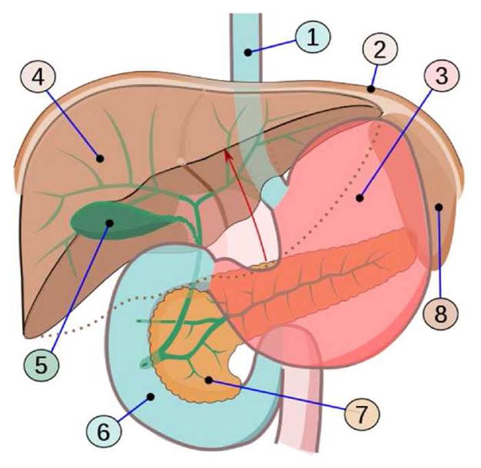
The lower border of the liver begins at the X intercostal space (spatium intercostale X) along the right midaxillary line (linea axillaris media dextra) and extends to the left along the margin of the right costal arch (arcus costalis dexter), crossing the IX costal Cartilage (cartilago costalis IX) on the right. Further, this border runs in the epigastric region (regio epigastrica) 1.5 cm below the xiphoid process (processus xiphoideus), crosses the VII costal cartilage (cartilago costalis VII) on the left, and reaches the V left intercostal space (spatium intercostale sinistrum V) at the parasternal line (linea parasternalis), which corresponds to the intersection point of the projections of the upper and lower borders of the liver (hepar).
Thus, only a small area of the liver surface (facies hepatis) in the epigastric region (regio epigastrica) is directly adjacent to the anterior abdominal wall.
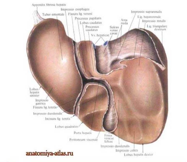
The liver is located in the Abdominal cavity, occupying the right hypochondriac region, partially the epigastric, and the left hypochondriac regions. The upper edge of the liver on the right runs along the 5th intercostal space, and on the left along the 6th intercostal space. The lower edge of the liver on the right runs along the margin of the costal arch (normally, the liver does not protrude from under the costal arch), and on the left, at the attachment site of the 8th left rib to the 7th. The liver features two surfaces: the upper convex (diaphragmatic) and the lower (visceral). The visceral surface has two longitudinal grooves—right and left—and one transverse groove; the gallbladder is located anteriorly within the right longitudinal groove. The liver consists of lobules, which are the Structural and functional units of the organ.
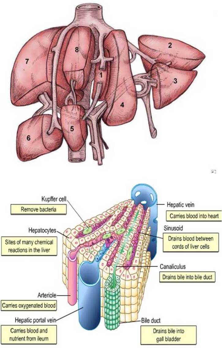
The lobules consist of liver Cells called hepatocytes. Hepatocytes secrete bile, which flows into ducts that initially merge into the right and left hepatic ducts, and then into a common hepatic duct that joins the cystic duct and empties into the duodenum. Blood enters the liver through two vessels: the portal vein, which brings blood from the unpaired abdominal Organs, and the hepatic artery, which supplies nutrients and oxygen; blood drains via the hepatic Veins into the INFERIOR VENA CAVA.
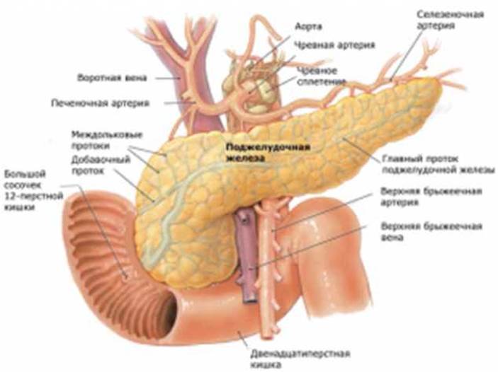
The pancreas is a gland of both internal and external secretion; it has a triangular shape and weighs 150 g. The pancreas is located in the abdominal cavity in the left hypochondriac region at the level of the 1st–2nd lumbar vertebrae. The Stomach is situated anterior to it, the HEAD of the pancreas is embraced by the duodenum, and the Spleen is located to the left. The gland consists of a head, tail, and body. The glandular cells secrete pancreatic juice, which enters the duodenum via ducts. Topography.
The structural and functional unit of The Endocrine Part of the Pancreas is the endocrine islets (islets of Langerhans, insulae pancreatici).
✵ The total number of islets ranges from 1 to 2 million.
✵ The diameter of an islet is 100–300 µm (for comparison, the size of an erythrocyte is 7 µm).
✵ Localization: among the acini (secretory units) of the exocrine part, separated from them by a thin layer of Connective Tissue.
✵ The largest number of islets is localized in the tail part of the gland. Basophilic cells (B cells, β-cells) make up the bulk of the islet cells (about 70–75%). Most of them lie in the center of the islets, though they may also be located near the capsule. These cells synthesize Insulin, which exerts a hypoglycemic effect by promoting the uptake of blood glucose by tissue cells.
Acidophilic cells (A cells, α-cells) make up 20–25% of the total cellular mass of the islets and are located in small groups throughout the islet. These cells produce the hormone Glucagon, which is an antagonist to insulin. Under its influence, enhanced Glycogenolysis (breakdown of glycogen to glucose) occurs in Tissues.
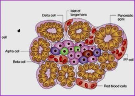
Dendritic cells (D cells) make up 5–10%, are located at the periphery of the islet, and produce Somatostatin, which inhibits Growth Hormone synthesis.
Argyrophilic cells (D1 cells) are found in small numbers within the islets and produce vasoactive intestinal peptide (VIP), which lowers blood pressure and stimulates the secretion of enzymes and hormones by the pancreas.
Last update: 08/08/2026
Editorial and Educational Adaptation: This material has been compiled based on the primary/original source text. The project team performed an editorial review, corrected technical inaccuracies, structured sections, and adapted the content for an educational format.
What was processed:
- elimination of formatting defects (OCR errors, structural breaks, corrupted characters);
- editorial organization of content;
- standardization of terminology in accordance with academic sources;
- verification of factual statements against the original source text.
All mentions of the author, publication year, and origin of the primary text have been preserved in accordance with the source.