Human Anatomy, Part 1 - K. A. Dyubenko, A. K. Kolomiyytsev, Yu. B. Chaykovsky 2002
Special Part
Splanchnology, splanchnologia [the study of viscera] — Development of the digestive and respiratory systems
Development of the intestinal tube
The appearance of the gut tube in the embryos of higher vertebrates is associated with The formation of body folds. By significantly reshaping the embryonic body through the tucking of its margins, these folds cause the embryonic (intestinal) endoderm and its associated regions, along with the visceral layers of the intraembryonic ventral mesoderm, to roll into a tube. These primordia ultimately form the primary gut of the embryo.
In a four-week-old embryo, the gut lies in the sagittal plane and follows a straight course, with the exception of a slight curvature caused by the bending of the entire body, as well as a flexure at the caudal end of the gut tube, which extends into the tail region of the embryo.
During the Formation of the gut, the visceral layers of the ventral mesoderm approach each other, forming the dorsal mesentery along the entire length of the gut tube, and the ventral mesentery in the region of The Stomach and the initial PARTS OF THE duodenum. The diameter of the gut tube is uniform throughout most of its length. Notable exceptions include the cranial part of the foregut, which participates in the formation of the Pharynx (Fig. 277A); a spindle-shaped dilatation representing the stomach primordium; and a slightly expanded region of the hindgut—the cloaca—from which the allantois develops (Fig. 277B).
The midgut does not immediately lose its connection with the yolk sac; instead, it communicates with the yolk endoderm via the yolk duct, which is incorporated into the yolk stalk. As noted previously, near the caudal region of the cloaca, an ectodermal invagination—the proctodeum—forms in the ventral body wall. Similar to the stomodeum region, the internal lining of the gut here sees the endoderm come into contact with the ectoderm, together forming a thin membrane. This membrane, known as the anal plate (or anal membrane), subsequently ruptures, thereby establishing the external opening of the hindgut.
The internal lining of the primary gut gives rise to the surface epithelium of the digestive tract mucosa and its associated glands. The other Tissues of the gastrointestinal tract (muscular and Connective Tissues) develop from the mesenchyme, which originates from the visceral layer of the ventral mesoderm. The latter also serves as the source for the simple squamous epithelium (mesothelium) that covers the serous membrane.
The Esophagus develops from a narrowed segment of the digestive tube located just inferior to the pharyngeal gut. Initially short, the esophagus elongates as the stomach shifts its position. Esophageal glands begin to appear by the fourth month, and due to the formation of longitudinal folds, its lumen acquires a stellate shape in cross-section. The esophageal epithelium is initially simple columnar; in the four-week-old embryo, it becomes stratified; by the third month, it transitions to pseudostratified ciliated epithelium; and from the sixth month onward, it is stratified squamous.
During the sixth and seventh weeks of Embryogenesis, both the shape and position of the stomach undergo significant changes driven by the following factors.
1. The gastric expansion grows longitudinally, with its posterior wall experiencing the most intense growth. As a result, the stomach primordium curves, forming the greater curvature located dorsally and the lesser curvature oriented ventrally.
2. With the formation of the greater curvature, the dorsal mesentery enlarges, bulging to the left and forming the primordium of the omental bursa (Fig. 277B).
3. The stomach rotates from left to right by approximately 90°. Consequently, the greater curvature, initially facing dorsally, comes to lie on the left side, while the lesser curvature, initially facing ventrally, occupies a right-sided position. The left wall of the stomach thus becomes anterior, and the right wall becomes posterior. This rotational movement extends to the lower section of the esophagus, which also rotates from left to right by approximately 90° (Fig. 277G). Because of this, the vagus nerves, which initially run along the sides of the esophagus, become asymmetrical: the left Vagus nerve shifts to the anterior surface of the stomach, while the right vagus nerve shifts to the posterior surface.
4. Due to the intensive growth of the Liver, the stomach, which previously had a vertical orientation, assumes an oblique-transverse position, while the dorsal mesentery bulges.
5. At approximately the seventh week of development, the stomach, along with The Heart and lung primordia, shifts inferiorly.
Class="center">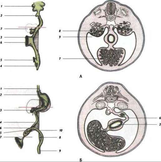
Fig. 277. Schematic representation of intestinal positioning and mesenteries during development:
A: 1 - pharynx; 2 - lung bud; 3 - dorsal mesentery; 4 - cloaca; 5 - allantoic duct; 6 - part of the yolk sac; 7 - liver primordium; 8 - mesonephros; 9 - peritoneal cavity; B: 1 - esophagus; 2 - stomach; 3 - Gallbladder; 4 - Small Intestine; 5 - cecum; 6 - mesentery; 7 - yolk stalk; 8 - mesentery of the Large Intestine; 9 - rectum; 10 - transverse colon.
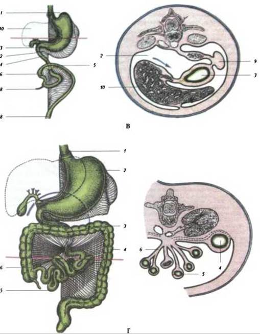
Fig. 277. Schematic representation of intestinal positioning and mesenteries during development (Continued):
B: 1 - esophagus; 2 - omental bursa; 3 - stomach; 4 - small intestine; 5 - transverse colon; 6 - ascending colon; 7 - vermiform Appendix; 9 - Spleen; 10 - liver; G: 1 - esophagus; 2 - stomach; 3 - transverse colon; 4 - descending colon; 5 - small intestine; 6 - mesentery of the small intestine
The gastric glands begin to form during the fourth month of embryonic development.
As noted previously, the primary gut initially appears as a straight tube extending through the entire body of the embryo. In its middle portion, it communicates with the yolk sac via the vitelline (omphalomesenteric) duct. Subsequently, during the sixth week of embryonic development, accelerated growth in the middle section of the gut gives rise to the primary intestinal loop, which lies in the sagittal plane and is seemingly drawn into the umbilical stalk by the vitelline duct.
Slightly caudal to the Water/144.html">Origin of the vitelline duct, a small thickening develops, serving as the primordium of the cecum. This expansion marks the boundary between the future small and large intestines.
Subsequently, the arrangement of the various segments of the gut becomes more complex due to growth and rotation. Viewed from an anterior perspective, the Rotation of the gut tube occurs from right to left, i.e., in a counterclockwise direction. In the process, the segment of the gut located caudal to the cecal primordium—corresponding to the colon—is seemingly "displaced" superiorly, coming to lie predominantly superior to the small intestine and crossing it. As it undergoes these changes, the large intestine assumes its definitive position and differentiates into the ascending, transverse, and descending colons. Elongation of the ascending colon begins in the fifth month and is completed only after fetal birth.
A fold forms between the cecum and the small intestine proper, which in turn gives rise to the ileocecal valve. The vermiform appendix develops from the cecum. Following the bending of the primary gut, the region destined to form the small intestine rapidly elongates. As a result, it proliferates and forms loops: initially three, and later seven. For a time, unable to fit within the Abdominal cavity—which is largely occupied by the liver—these small intestinal loops herniate outside the cavity and enter the umbilical cord primordium. The resulting ventral body wall herniation was termed the physiological umbilical hernia. By the tenth week, owing to the increasing volume of the abdominal cavity, the intestinal loops retract into it and assume their definitive position, localizing primarily in the left side of the abdominal cavity.
The rotation of the gut is part of a broader rotational process affecting the entire gut tube, extending to the stomach and the lower esophagus. Due to the rotation of the stomach, the adjacent segment corresponding to the duodenum is brought close to the posterior abdominal wall and becomes fixed in place.
As loops form, the dorsal mesentery of the small intestine acquires a folded Structure, which determined its terminology (mesentery originating from medieval garments featuring ruffled collars worn in several Western European countries).
The vitelline duct obliterates concurrently with the closure of the ventral wall of the intestine and the ventral body wall. Occasionally, its proximal part persists, forming the so-called Meckel's diverticulum, which extends from the ileum.
As noted, the most caudally located section of the intestinal tube expands to form the cloaca, with the allantois originating from its anterior aspect. The embryonic URINARY AND REPRODUCTIVE tracts open into the cloacal cavity. The cloaca is divided into an anterior part (the urogenital sinus) and a posterior part (the rectum) by a frontally positioned urorectal septum, which consists of mesenchyme and develops near THE ORIGIN OF the allantois. As the name implies, the latter part gives rise to the rectum.
Upon reaching the cloacal membrane, the urorectal septum divides it into an anterior urogenital membrane and a posterior anal membrane. These membranes subsequently rupture, giving rise to the respective orifices. The caudal section of the urorectal septum, which separates these openings, serves as the primordium of the Perineum.
The ectoderm lining the depression (proctodeum)—formed at the site where the hindgut ectoderm contacts the cutaneous ectoderm—invaginates inward to join the endoderm. This process leads to the formation of the anal pit and a secondary anal opening lined with epithelium of an ectodermal type.
Occasionally, the anal membrane fails to rupture, resulting in an imperforate rectum lacking an external opening. This developmental anomaly, known as atresia ani, requires surgical intervention.
The epithelium of the digestive tract and its associated wall glands develops from the endoderm of the primitive gut. An exception to this is the foregut, whose epithelial lining is only formally of endodermal origin. In fact, the epithelial component of the pharyngeal gut and its respiratory derivatives exhibits structural features and properties characteristic of epidermal-type epithelia. A concrete example of this is the transition zone between the esophagus and the stomach, where two genetically distinct epithelia meet: the ectodermal epithelium characteristic of the Oral Cavity, pharynx, and esophagus, and the endodermal epithelium that constitutes the lining of the small intestine and the greater part of the large intestine. In lower vertebrates and reptiles, the material lining the foregut invaginates during Gastrulation; however, this phenomenon has not been observed in birds and mammals.
The Connective Tissue and smooth Muscle elements of the digestive tract develop from the mesenchyme. Nerve fibers and migrating neuroblasts grow into the intestinal wall via the dorsal mesentery, leading to the formation of the nervous apparatus of the digestive Organs. The primary Developmental anomalies of the intestinal tube include atresia, Meckel's diverticulum, cardiospasm, and Hirschsprung's disease.
Last update: 08/08/2026
Editorial and Educational Adaptation: This material has been compiled based on the primary/original source text. The project team performed an editorial review, corrected technical inaccuracies, structured sections, and adapted the content for an educational format.
What was processed:
- elimination of formatting defects (OCR errors, structural breaks, corrupted characters);
- editorial organization of content;
- standardization of terminology in accordance with academic sources;
- verification of factual statements against the original source text.
All mentions of the author, publication year, and origin of the primary text have been preserved in accordance with the source.