Human Anatomy Part 1 - K. A. Dyubenko, A. K. Kolomiyytsev, Y. B. Chaykovsky 2002
Special Part
Splanchnology, splanchnologia [the study of viscera] — Development of the digestive and respiratory systems
Development of the liver
The Liver begins to develop at the end of the third week of embryonic life. The source of its epithelial development is the entoderm of the part of the gut tube that corresponds to the future duodenum. Here, an outgrowth appears (the hepatic diverticulum) (Fig. 278), which grows into the mesenchyme of the septum transversum formed by the ventral body wall; it grows in a dorsal direction and serves as one of the primordia of the Diaphragm. This outgrowth also penetrates the ventral mesentery. In the distal PARTS OF THE diverticulum, cords of hepatic Cells are formed, representing the secretory region of the liver (hepatic cords or trabeculae), while the proximal parts give rise to the hepatic ducts. At the junction of the ducts, a dilation forms, which represents the primordium of the Gallbladder, and near the gut, the diverticulum forms an outpocketing that serves as the ventral primordium of the Pancreas.
The hepatic cords come into contact with wide (sinusoidal) capillaries. In the postnatal period, they receive nutrient-rich Blood from the intestines via the portal vein. In the prenatal period, these substances reach the liver via the umbilical vein.
A significant portion of the stalk connecting the hepatic diverticulum to the duodenum elongates to form the common Bile duct, ductus choledochus, the cystic duct, ductus cysticus, and the common hepatic duct, ductus hepaticus communis.
The epithelial Components of the liver, gallbladder, and ducts develop from the entoderm. Their connective and Smooth Muscle Tissues differentiate from the mesenchyme. The peritoneal covering of the liver arises from the ventral mesentery. During the Embryonic period, the liver also performs Hematopoietic and Immune Functions.
Following the final formation of its primordium, the liver grows rapidly, thus becoming a large organ. By the end of the third month, its inferior margin reaches the inguinal region of the embryo. Subsequently, the growth of the liver slows down, and at birth, its lower margin lies at the level of the umbilicus. In an adult, it is located even higher.
Class="center">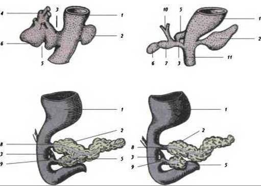
Fig. 278. Diagram of The Development of The Liver and pancreas (after J. Stieve):
1 - Stomach; 2 - dorsal pancreas; 3 - bile duct; 4 - liver primordium; 5 - ventral pancreas; 6 - gallbladder primordium; 7 - cystic duct; 8 - minor pancreatic duct; 9 - major pancreatic duct (duct of Wirsung); 10 - hepatic duct; 11 - duodenum
Last update: 08/08/2026
Editorial and Educational Adaptation: This material has been compiled based on the primary/original source text. The project team performed an editorial review, corrected technical inaccuracies, structured sections, and adapted the content for an educational format.
What was processed:
- elimination of formatting defects (OCR errors, structural breaks, corrupted characters);
- editorial organization of content;
- standardization of terminology in accordance with academic sources;
- verification of factual statements against the original source text.
All mentions of the author, publication year, and origin of the primary text have been preserved in accordance with the source.