Human Anatomy, Part 1 - K. A. Dyubenko, A. K. Kolomiytsev, Yu. B. Chaykovsky 2002
Special Part
Arthrology, artrologia [the study of bone joints] - General Overview
Continuous bone joints - synarthroses
Continuous bone joints are earlier in development. They are characterized by considerable strength, low flexibility, high elasticity, and limited movement. Depending on The Structure of the connecting tissue, continuous bone joints are divided into Three types of synarthroses (BNA).
1. Fibrous joints, junctura fibrosa s. syndesmosis.
2. Cartilaginous joints, junctura cartilaginea s. synchondrosis.
3. Bony joints, junctura ossea s. synostosis.
Fibrous joints are formed after birth when Connective Tissue remains between the bones, providing bone connection.
1. Fibrous joints (syndesmoses) include: interosseous membranes, membranae interosseae, ligaments, ligamenta (Fig. 75B), cranial sutures, suturae cranii, gomphoses, and fontanelles, fonticuli.
Class="center">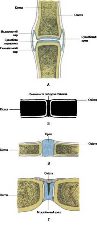
Fig. 75. Types of bone joints:
A - joint (articular); B - syndesmosis; C - synchondrosis; D - symphysis (hemiarthrosis)
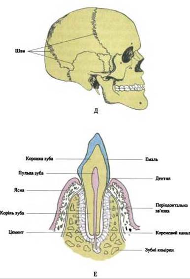
Fig. 75. Types of bone joints (Continued)
E - cranial sutures; F - gomphosis
Interosseous fibrous membranes, membranae interosseae fibrosae, connect adjacent bones. They are located between the BONES OF THE forearm, membrana interossea antebrachii, and between the bones of the leg, membranae interosseae cruris, or close openings in bones, such as the obturator membrane, membrana obturatoria, and the anterior and posterior atlanto-occipital membranes, membranae atlantooccipitales anterior et posterior. Interosseous membranes connect bones and provide a larger surface area for Muscle attachment. They are formed mainly by bundles of Collagen fibers and feature openings for the passage of Blood Vessels and nerves.
Ligaments, ligamenta, serve to secure bone connections. They can be very short, such as the dorsal intercarpal ligaments, ligg. intercarpalia dorsalia, or, conversely, long, such as the anterior and posterior longitudinal ligaments of THE Vertebral Column, ligg. longitudinale anterius et posterius.
Ligaments are strong fibrous cords consisting of longitudinal, oblique, and intersecting bundles of collagen fibers with a small number of elastic fibers. Ligaments can withstand high tensile loads. This group also includes ligaments formed exclusively by elastic fibers. They are not as strong as fibrous syndesmoses, but are highly stretchable and flexible—these are the yellow ligaments, ligamenta flava, located between the vertebral arches.
Cranial sutures, suturae cranii (Fig. 75D), occur only in The Skull and are a type of syndesmosis in which the bone edges are firmly joined by thin layers of Fibrous connective tissue. Sutures are characterized by exceptional strength. Depending on the shape of the skull bones, the following types of sutures are distinguished:
- serrate suture, sutura serrata s. dentata (BNA), in which the edge of one bone has projections that fit into depressions In the second bone (e.g., the frontoparietal suture);
- squamous suture, sutura squamosa, characterized by the bevelled, scale-like edge of one bone overlapping the bevelled edge of another (e.g., the temporoparietal suture);
- plane suture, sutura plana s. harmonia (BNA), in which the smooth edge of one bone apposes a similar edge of another without forming projections, typical of facial skull bones (e.g., between the nasal bones).
Gomphosis is a type of fibrous bone joint. It can be observed between the roots of the Teeth and the dental alveoli (dentoalveolar syndesmosis, syndesmosis dentoalveolaris) (Fig. 75E). A connective tissue layer, the periodontium, is located between the tooth and the Bone tissue of the socket.
2. In cartilaginous joints (synchondroses), bones are connected by a layer of fibrocartilage or hyaline Cartilage (Fig. 75C). Hyaline cartilage harmoniously combines strength and elasticity. Synchondroses are quite strong and resilient, thereby performing Shock-absorbing Functions. The mobility of this joint is insignificant and depends on the thickness of the cartilaginous layer—the greater the thickness, the greater the mobility, and vice versa. An example of synchondroses formed by fibrocartilage is the intervertebral discs, disci intervertebrales, located between the vertebral bodies. They are strong and resilient, acting as buffers against jolts and shocks. An example of synchondroses formed by hyaline cartilage is the epiphyseal plates located at the border between epiphyses and metaphyses in long bones, or the costal cartilages connecting the Ribs to the Sternum. Depending on their duration, synchondroses can be temporary, existing up to a certain age (e.g., the cartilaginous junctions between the diaphysis and epiphyses of long bones and the three pelvic bones), or permanent, persisting throughout human life (e.g., between the pyramid of the Temporal bone and adjacent bones: the sphenoid and occipital). A variation of synchondrosis is the Pubic Symphysis, symphysis pubica, where bones are also connected by cartilage containing a small cavity.
3. If a temporary continuous joint (fibrous or cartilaginous) is replaced by bone tissue, it is called a synostosis (BNA). This type of joint is the strongest, but it loses its elastic function. Examples of synostoses in adults include the fusion between the bodies of the occipital and sphenoid bones, among the sacral vertebrae, and between the PARTS OF THE Mandible.
Last update: 08/08/2026
Editorial and Educational Adaptation: This material has been compiled based on the primary/original source text. The project team performed an editorial review, corrected technical inaccuracies, structured sections, and adapted the content for an educational format.
What was processed:
- elimination of formatting defects (OCR errors, structural breaks, corrupted characters);
- editorial organization of content;
- standardization of terminology in accordance with academic sources;
- verification of factual statements against the original source text.
All mentions of the author, publication year, and origin of the primary text have been preserved in accordance with the source.