HUMAN MEDICAL BIOLOGY, ANATOMY, PHYSIOLOGY AND PATHOLOGY - Y.I. Fedoniuk 2010
ANATOMY, PHYSIOLOGY, PATHOLOGY
CHAPTER 3. ANATOMICAL AND PHYSIOLOGICAL ASPECTS OF BODY FUNCTION SELF-REGULATION
AUTONOMIC DIVISION OF THE PERIPHERAL NERVOUS SYSTEM
ANATOMICAL STRUCTURE OF THE AUTONOMIC DIVISION OF THE PERIPHERAL NERVOUS SYSTEM
1. SYMPATHETIC DIVISION
The Sympathetic division of the Autonomic Nervous system consists of central and peripheral parts. The central part is formed by the Cells of the lateral horns of the Spinal Cord at the level of the eighth cervical, all thoracic, and the upper three lumbar segments. The peripheral part comprises nerve fibers and sympathetic ganglia. A distinction is made between paravertebral (lateral) and prevertebral ganglia. Paravertebral ganglia are connected by interganglionic fibers to form the right and left sympathetic trunks.
A characteristic feature of postganglionic sympathetic fibers is The formation of plexuses along the course of the Arteries that supply a given organ.
The Sympathetic trunk is a chain of paravertebral ganglia interconnected by interganglionic fibers. The right and left trunks are located laterally along THE Vertebral Column (Fig. 3.52). White rami communicantes (preganglionic fibers) from the Spinal Nerves approach the ganglia of the sympathetic trunk. Gray rami communicantes (postganglionic fibers) pass from all the ganglia of the sympathetic trunk to the peripheral spinal nerves. The sympathetic trunk is subdivided into cervical, thoracic, lumbar, sacral, and coccygeal parts.
The cervical part is represented by superior, middle, and inferior ganglia. The superior cervical ganglion is the largest, while the inferior ganglion often fuses with the first thoracic ganglion to form the stellate ganglion. Branches innervating the Organs of the HEAD and Neck depart from the superior cervical ganglion. These branches form plexuses on the internal and external carotid arteries and, together with their branches, reach the lacrimal gland, Salivary Glands, Glands of the mucous membrane of the Pharynx, Larynx, Tongue, and the pupil dilator Muscle. Cardiac nerves emerge from all three ganglia; in the thoracic cavity, together with the Vagus nerve, they form the superficial and deep cardiac plexuses, from which nerves extend to The Heart wall.
Class="center">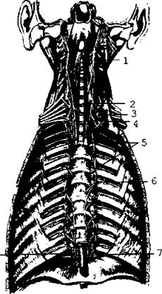
Fig. 3.52. Cervical and thoracic PARTS OF THE sympathetic trunk; anterior view
The thoracic part consists of 10-12 ganglia, from which branches extend to the aorta, heart, Lungs, Bronchi, and Esophagus. The greater and lesser splanchnic nerves are the largest nerves originating from the V-IX and X-XI thoracic ganglia. They pass through the Diaphragm into the Abdominal cavity AND participate in the Formation of the abdominal (celiac) plexus.
The lumbar part consists of 3-5 ganglia. They give rise to branches that form the autonomic nerve plexuses of the abdominal cavity and pelvis (Fig. 3.53).
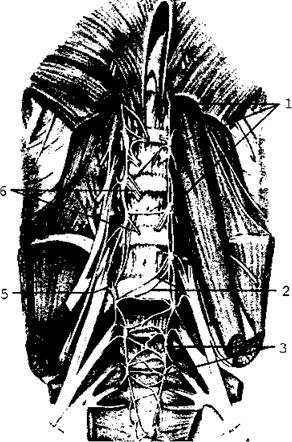
Fig. 3.53. Lumbar and sacral parts of the sympathetic trunk: 1 - lumbar ganglia; 2 - rami communicantes (lumbar); 3 - sacral ganglia; 4 - ganglion impar; 5 - ramus communicans (gray); 6 - lumbar splanchnic nerves, greater and lesser splanchnic nerves.
The sacral part consists of four ganglia located on the anterior surface of the sacrum. Inferiorly, the ganglion chains of the left and right sympathetic trunks converge at a single unpaired coccygeal ganglion (ganglion impar). All these structures are integrated into the pelvic part of the sympathetic trunks.
Branches extending from them form the Autonomic plexuses of the pelvis.
Last update: 08/08/2026
Editorial and Educational Adaptation: This material has been compiled based on the primary/original source text. The project team performed an editorial review, corrected technical inaccuracies, structured sections, and adapted the content for an educational format.
What was processed:
- elimination of formatting defects (OCR errors, structural breaks, corrupted characters);
- editorial organization of content;
- standardization of terminology in accordance with academic sources;
- verification of factual statements against the original source text.
All mentions of the author, publication year, and origin of the primary text have been preserved in accordance with the source.