HUMAN MEDICAL BIOLOGY, ANATOMY, PHYSIOLOGY AND PATHOLOGY - Y.I. Fedoniuk 2010
ANATOMY, PHYSIOLOGY, PATHOLOGY
CHAPTER 3. ANATOMICAL AND PHYSIOLOGICAL ASPECTS OF BODY FUNCTION AUTO-REGULATION
AUTONOMIC DIVISION OF THE PERIPHERAL NERVOUS SYSTEM
ANATOMICAL STRUCTURE OF THE AUTONOMIC DIVISION OF THE PERIPHERAL NERVOUS SYSTEM
2. PARASYMPATHETIC DIVISION
The Parasympathetic division of the Autonomic Nervous system also comprises central and peripheral parts.
The central part is represented by parasympathetic nuclei located in the Midbrain, Hindbrain (Pons and Medulla Oblongata), and the sacral segments of the Spinal Cord.
The peripheral part consists of ganglia and fibers that are Components of the III, VII, IX, and X cranial nerve pairs, as well as the pelvic nerves.
In the midbrain, adjacent to the motor Nucleus of the third cranial nerve pair, lies the parasympathetic accessory nucleus (Edinger-Westphal nucleus), whose Cell processes contribute to the Oculomotor nerve.
They synapse in the ciliary ganglion, located within the Orbit, and innervate the sphincter pupillae Muscle and the ciliary muscle.
In the Rhomboid fossa, next to the Facial Nerve nucleus, lies the superior salivatory nucleus. Its cell processes belong to the intermediate nerve, which runs within the facial nerve.
Together with Branches of the facial and trigeminal nerves, postganglionic parasympathetic fibers enter the lacrimal gland, the mucous Glands of the nasal and oral cavities, as well as the submandibular and sublingual Salivary Glands. These fibers synapse in the pterygopalatine, submandibular, and sublingual ganglia.
Parasympathetic fibers from the inferior salivatory nucleus travel within the IX cranial nerve pair to the Cytology/practical/97.html">Parotid salivary gland. These fibers synapse in the otic ganglion.
The largest number of parasympathetic fibers runs within the Vagus nerve. Originating from its dorsal parasympathetic nucleus, they innervate all Organs of the neck, thoracic, and abdominal cavities down to the transverse colon. In the thoracic and abdominal cavities, the vagus nerve fibers approach the sympathetic plexuses and enter the organs alongside them. The parasympathetic fibers of the vagus nerve synapse in intramural ganglia (located within the organ walls).
The descending and sigmoid colons, as well as all pelvic organs, receive their parasympathetic fibers from the pelvic splanchnic nerves branching off the sacral spinal cord. These form the autonomic nerve plexuses of the pelvis and synapse in the ganglia of the pelvic organ plexuses.
Within the thoracic, abdominal, and pelvic cavities, sympathetic and parasympathetic fibers form mixed plexuses. The largest plexus in the Abdominal cavity is the celiac (solar) plexus, located on the anterior surface of the Abdominal Aorta (Fig. 3.54). It includes two celiac ganglia and the superior mesenteric ganglion. Preganglionic fibers are carried to them by Postganglionic fibers form plexuses along Blood Vessels and spread in all directions toward the organs. Branches of the vagus nerves also approach the celiac plexus, proceeding to the organs as part of the solar plexus.
Class="center">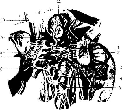
Fig. 3.54. Celiac plexus: 1 - left celiac ganglion; 2 - suprarenal plexus; 3 - left aortico-renal ganglion; 4 - superior mesenteric ganglion; 5 - intermesenteric plexus; 6 - left aortico-renal ganglion; 7 - renal plexus; 8 - right celiac ganglion; 9 - posterior vagal trunk; 10 - greater splanchnic nerve; 11 - anterior vagal trunk.
A rather large autonomic nerve plexus extends downward along the abdominal aorta, forming the superior hypogastric plexus in the region of the sacral promontory. It descends into the pelvic cavity and continues into the right and left inferior hypogastric plexuses, which are joined by the pelvic parasympathetic nerves.
Interlacing near each pelvic organ, all these branches form plexuses that provide their innervation.
Last update: 08/08/2026
Editorial and Educational Adaptation: This material has been compiled based on the primary/original source text. The project team performed an editorial review, corrected technical inaccuracies, structured sections, and adapted the content for an educational format.
What was processed:
- elimination of formatting defects (OCR errors, structural breaks, corrupted characters);
- editorial organization of content;
- standardization of terminology in accordance with academic sources;
- verification of factual statements against the original source text.
All mentions of the author, publication year, and origin of the primary text have been preserved in accordance with the source.