MEDICAL BIOLOGY, HUMAN ANATOMY, PHYSIOLOGY AND PATHOLOGY - Ya.I. Fedoniuk 2010
BIOLOGY
CHAPTER 1. BIOLOGICAL BASES OF HUMAN VITAL ACTIVITY
1.2. CELL. TISSUES. CONCEPT OF ORGAN, ORGAN SYSTEM, ORGANISM
EPITHELIAL TISSUE
2. Glandular Epithelium
Glandular epithelium consists of glandular Cells, or glandulocytes. These are specialized cells adapted to the synthesis, accumulation, and secretion of substances.
Glands are subdivided into Endocrine (Ductless) Glands and exocrine glands; accordingly, glandulocytes are classified into endocrinocytes and exocrinocytes. Endocrine glands form the Endocrine System, which, together with The Nervous system, regulates and coordinates the functioning of Organs throughout the body. Their characteristic feature is that they release their secretions (Hormones) directly into the bloodstream. These glands include the Pituitary Gland, Pineal Gland, thyroid and Parathyroid glands, Adrenal Glands, as well as the endocrine PARTS OF THE Pancreas and Gonads.
Exocrine glands possess terminal secretory portions and excretory ducts. They release their secretions into cavities (for example, the gastric cavity) or onto The surface of an epithelial layer.
Depending on the number of cells, all body glands are divided into unicellular and multicellular. The latter are present in the body in large numbers.
Based on their Location (relative to the epithelial layer), endoepithelial and exoepithelial glands are distinguished, meaning those located within the epithelium or outside it, respectively. The majority of glands are exoepithelial (Fig. 1.5).
Class="center">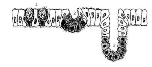
Fig. 1.5. Various types of exocrine glands: 1 - unicellular endoepithelial glands (goblet cells); 2 - multicellular endoepithelial gland (of the nasal mucosa); 3 - multicellular exoepithelial gland.
Exocrine glands differ from one another in Structure, mode of secretion, and COMPOSITION OF THE secretion. Based on The structure of their terminal portions and excretory ducts, several types are distinguished (Fig. 1.6).
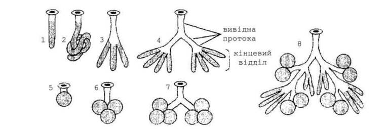
Fig. 1.6. Morphological Classification of exocrine glands According to the structure of their terminal portions and excretory ducts: 1 - simple unbranched tubular gland; 2 - simple unbranched tubular gland with a coiled terminal portion; 3 - simple branched tubular gland; 4 - compound branched tubular gland; 5 - simple unbranched alveolar gland; 6 - simple branched alveolar gland; 7 - compound branched alveolar gland; 8 - compound branched tubulo-alveolar gland.
A simple gland has an unbranched excretory duct, which distinguishes it from a compound gland whose excretory duct is branched. To differentiate between a branched and an unbranched gland, one must examine its secretory portion. A branched gland always has multiple terminal portions. According to the shape of their secretory units, glands are classified into tubular, alveolar, and tubulo-alveolar.
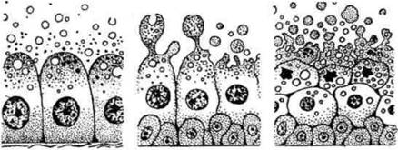
According to the mode of secretion release from The Cell, glands are divided into three types: merocrine, apocrine, and holocrine (Fig. 1.7). In merocrine secretion, glandular cells are not destroyed. Merocrine glands include the Salivary Glands, pancreas, the majority of Sweat Glands, etc. In apocrine secretion, the apical part of the cell detaches along with the secretion. Examples of apocrine glands are the Mammary Glands and the sweat Glands of the axillary regions. In holocrine secretion, the entire cell is destroyed. An example of a holocrine gland is the sebaceous gland of the Skin.
According to The chemical composition of their secretion, mucosal, serous (protein), mixed (seromucous), sweat, and Sebaceous Glands are distinguished.
a b c
Fig. 1.7. Schematic representation of secretion types: a - merocrine; b - apocrine; c - holocrine.
Last update: 08/08/2026
Editorial and Educational Adaptation: This material has been compiled based on the primary/original source text. The project team performed an editorial review, corrected technical inaccuracies, structured sections, and adapted the content for an educational format.
What was processed:
- elimination of formatting defects (OCR errors, structural breaks, corrupted characters);
- editorial organization of content;
- standardization of terminology in accordance with academic sources;
- verification of factual statements against the original source text.
All mentions of the author, publication year, and origin of the primary text have been preserved in accordance with the source.