Nephrology for the General Practitioner - O.I. Bakaliuk 2003
Emergency Uro-Nephrology
Acute Urinary Retention
Therapists, especially general practitioners, frequently have to provide emergency uro-nephrological care. The patient's prognosis for recovery or even survival largely depends on choosing the correct medical strategy, particularly in the absence of a urologist or nephrologist. Therefore, we consider it appropriate to highlight key aspects of this management strategy for the most common clinical scenarios. A detailed coverage of emergency uro-nephrological care can be found in the monographs by Yu.A. Pytel et al. (1985), O.V. Lyulko et al. (1996), E.V. Shakhov (1998), and I.B. Osipov et al. (1999).
The inability to urinate voluntarily despite a distended bladder and the urge to urinate is termed acute urinary retention. Anamnesis helps determine the underlying cause of this pathological condition: establishing the voiding pattern, the volume and visual characteristics of urine prior to retention, the precipitating factors, as well as the presence and course of concomitant diseases, injuries, etc.
The most common causes of acute urinary retention include:
- impaired Innervation of the bladder, sphincters, and Urethra due to trauma, metastatic tumors, or lumbar spine fractures;
- traumatic injuries to the bladder and urethra;
- mechanical obstructions along the Urinary Tract;
- venous congestion in the pelvic Organs accompanied by their engorgement, particularly the Prostate Gland (resulting from a sedentary lifestyle, constipation, cold exposure of the feet, forced delay of urination, alcohol consumption, or spicy food);
- Psychogenic Disorders (fright, hysteria) and neurogenic bladder dysfunction, especially in children;
- postoperative states (forced horizontal position leading to venous congestion, wound pain upon straining the anterior abdominal wall Muscles, decreased detrusor tone following general anesthesia or spinal anesthesia, and the absence of the Micturition reflex in the supine position, etc.).
The most frequent causes of urinary retention in men are shown in Figure 103.
In women, acute urinary retention can also be caused by Colles' syndrome.
A distinct entity is acute urinary retention of the paradoxical ischuria type, where urine drips spontaneously from an overdistended bladder, unlike true Urinary Incontinence, in which urine leaks out immediately upon entering the bladder. As a rule, paradoxical ischuria develops gradually (e.g., against the Background of prostatic hyperplasia), although it may also present acutely (in Spinal Cord trauma or tumors).
Class="center">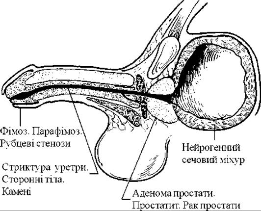
Fig. 103. Most frequent causes of urinary retention in men.
The Clinical presentation of acute urinary retention is characteristic: urges to urinate, a bursting pain above the pubic bone, in the Perineum, and the rectum. Patients are restless, assume various positions in an attempt to empty the bladder, press on the suprapubic area, etc. Physical examination reveals a suprapubic bulge, the upper limit of which may even reach the level of the umbilicus.
Palpation of this mass is quite painful.
Emergency care for acute urinary retention consists of immediate bladder decompression. This is achieved through various approaches: medication, catheterization of the bladder, suprapubic puncture, or cystostomy tube placement.
Reflex measures and pharmacotherapy are most commonly prescribed for postoperative urinary retention. If the patient's condition permits, a vertical posture and a heating pad applied to the suprapubic area are used. Antispasmodics are employed to relieve spasms of the bladder sphincter muscles (see: Emergency Management of Renal Colic). When bladder Muscle atony is the cause of acute urinary retention, one may administer neostigmine (subcutaneously, 1 ml of a 0.05% solution), pilocarpine (subcutaneously, 1 ml of a 1% solution), methenamine (intravenously, 5–10 ml of a 40% solution), or potassium acetate (orally, ex 60.0 : 300.0 — 1 tablespoon every 1–2 hours). Herbal remedies include aloe juice (1 teaspoon 2–3 times a day before meals), wild angelica ROOT decoction (ex 20.0 : 200.0 — 1 Glass three times a day), and fresh gooseberry or red currant fruits.
Soft catheters are preferred for bladder catheterization. The inability to catheterize the bladder using either soft or metal catheters provides the indication for capillary puncture, suprapubic cystostomy, or puncture cystostomy.
In clinical practice, bladder catheterization with a soft catheter or capillary bladder puncture is most frequently utilized.
Technique for inserting a soft catheter in men. The patient lies supine with legs slightly abducted. The physician stands to the patient's right. The glans Penis and the external urethral meatus are treated with an antiseptic solution and wiped dry. Using the third and fourth fingers of the left hand, the physician grasps the glans penis near the corona glandis and pulls it slightly forward and upward (Fig. 104).
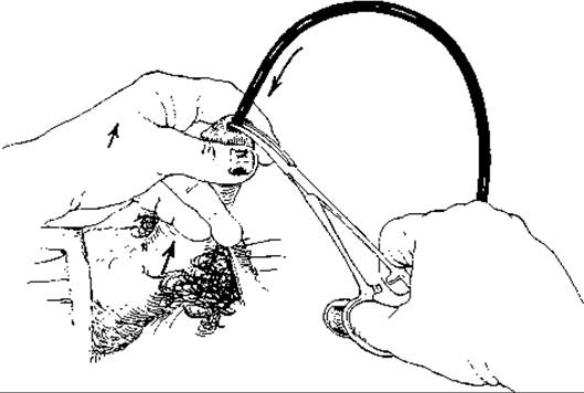
Fig. 104. Technique of bladder catheterization with a soft catheter in men.
The thumb and index fingers of the same hand spread the Lips of the external urethral meatus. The tip of the soft catheter is lubricated with sterile glycerin or petrolatum jelly. Holding it between the fourth and fifth fingers of the RIGHT HAND AND using forceps, the catheter is smoothly and gently advanced into the urethra without excessive force. The appearance of urine from the catheter indicates the Procedure's success.
Technique for inserting a soft catheter in women. This procedure is much simpler in women than in men. The patient lies supine with her hips and knees flexed (preferably in a gynecological chair). Following antiseptic preparation of the external urethral meatus and the vaginal introitus, the catheter (rubber or metal) is smoothly advanced into the bladder. The outer end of the catheter is secured to the Skin of the thigh with gauze strips.
Catheterization of the Urinary Bladder in girls and boys is practically identical to the procedure described for men and women, with the exception of using special (pediatric-sized) catheters. In girls, metal catheters are preferred.
Capillary puncture of the urinary bladder is performed along the midline of the abdomen, 1-2 cm superior to the Pubic Symphysis after determining the upper border of the bladder by Percussion. In this situation, the Peritoneum is displaced upward, allowing the extraperitoneal portion of the bladder wall to approach the anterior abdominal wall.
Procedure for capillary puncture of the urinary bladder. With the patient in the supine position, the abdominal wall is punctured strictly perpendicular to the skin, 2-3 cm above the pubic bone, using a 15-20 cm long needle with a stylet. The needle is gradually advanced deeper until it enters the bladder cavity. The criterion for correct needle placement is the outflow of urine after removing the stylet (Fig. 105).
A rubber tube is attached to the needle hub, and the urinary bladder is emptied. To prevent urine leakage into the preprostatic cellular tissue upon needle withdrawal, the soft Tissues surrounding the needle should be tightly compressed.
All other interventions for acute urinary retention (such as metal catheterization, urethral bougienage, trocar suprapubic cystostomy, standard cystostomy, transvesical suprapubic prostatectomy, etc.) must be performed in a specialized urology department.
Last update: 08/08/2026
Editorial and Educational Adaptation: This material has been compiled based on the primary/original source text. The project team performed an editorial review, corrected technical inaccuracies, structured sections, and adapted the content for an educational format.
What was processed:
- elimination of formatting defects (OCR errors, structural breaks, corrupted characters);
- editorial organization of content;
- standardization of terminology in accordance with academic sources;
- verification of factual statements against the original source text.
All mentions of the author, publication year, and origin of the primary text have been preserved in accordance with the source.