Botany - B.E. Yakubenko 2017
Part One. Plant Anatomy and Morphology
Chapter I. Cytology
Laboratory Class Topic 2.4. Reserve Nutrients
General Remarks. During metabolic processes, Cells produce various reserve nutrients. Most commonly, these are Proteins, CARBOHYDRATES, and Lipids. During Photosynthesis, glucose undergoes polymerization to form assimilatory or primary starch, which accumulates in the leaves. In storage Tissues, secondary or reserve starch is formed in the shape of starch grains. The Organelles responsible for starch synthesis in The Cell are amyloplasts, which are specialized leukoplasts that synthesize carbohydrates. Leukoplasts that store proteins are called proteinoplasts; those accumulating lipids are termed oleoplasts, and those storing Water or salt solutions are referred to hydrosol/hydroplasts.
Because starch deposition in potato tubers or wheat grains occurs unevenly throughout the day, a distinct stratification is visible in them. Starch layers deposited during daylight hours are loose, dark, and more saturated with hygroscopic moisture. Conversely, layers formed at night are narrow, light-colored, and dense. They are laid down as glucose arrives, concentrating around the center of formation (hilum). There are Three types of starch grains: simple, compound, and semi-compound. The first two exhibit individual stratification, whereas semi-compound grains possess both individual and common stratification encompassing several simple grains.
Reserve protein is deposited in three forms: 1) as aleurone grains, formed by the desiccation of vacuoles that contained water-soluble protein; 2) as gluten, which is deposited in the starchy part of the endosperm and contains up to 90% protein; 3) as crystalloids, found in potato tubers.
1. Common wheat grain (Triticum aestivum L.)
2. Potato tuber (Solanum tuberosum L.)
3. Castor bean seed (Ricinus communis L.)
4. Cultivated oat grain (Avena sativa L.)
5. Common buckwheat fruit (Fagopyrum esculentum Moench.)
Tasks:
1. Using a prepared slide of a wheat grain transverse section of the endosperm, examine its Structure and the reserve nutrients produced by the cell.
2. Using a freshly prepared slide of a potato tuber, examine the types of starch grains and their structure.
3. Using a freshly prepared slide of a castor bean seed, examine its structure and the types of aleurone grains.
4. Using freshly prepared slides of an oat grain and a buckwheat achene, examine the Structural Features of starch grains.
5. Sketch the observed objects in your laboratory notebook and provide the necessary labels.
Equipment and Materials: MBR-1 or Biolam microscopes, prepared slides, distributed plant material, Reagents, scalpels, razors, forceps, blades, wall charts, etc.
Microscopic examination of the common wheat grain slide. Under low magnification, examine The structure of the wheat grain and sketch the distribution of its individual structures schematically in your notebook. Then, examine these structures in greater detail using high magnification.
The first of these structures is the pericarp. Directly beneath it lies a dense, thin layer of the seed coat (testa), which is fused with the pericarp along its entire perimeter—a characteristic feature of the cereal fruit, known as a caryopsis.
The second characteristic STRUCTURE OF THE wheat grain is the aleurone layer, formed by nearly rectangular cells. Each of these cells contains A large number of aleurone grains. In addition to aleurone grains, locate the somewhat larger Cell Nucleus.
The third characteristic structure of the grain is the starchy endosperm. It occupies the major part of the grain and is formed by large, polygonal cells separated in places by intercellular spaces. This entire tissue has a light blue color and is easily recognizable. The cells of this endosperm region are packed with large and small starch grains exhibiting concentric stratification, interspersed with layers of gluten (Fig. 18).
|
Class="center"> |
Figure 18. Part of a transverse section of a rye grain: 1 — pericarp; 2 — intermediate layer; 3 — chlorophyll-bearing layer; 4 — integument (seed coat); 5 — remnants of nucellus; 6 — aleurone layer; 7 — aleurone grains; 8 — endosperm; 9 — starch grains |

Compare the observed slide with the wall chart, clarify the structural features of individual elements, and record your findings in your laboratory notebook.
Method for preparing a slide of potato starch grains. Take a Glass Microscope slide and a coverslip, and wipe them clean and dry. Place a drop of a weak iodine-potassium iodide solution on the slide and a drop of water right next to it. Gently rub a piece of sliced potato in both drops to release the starch grains. As a result, you will obtain a uniform, turbid suspension in the water drop. Cover this suspension with a coverslip. Repeat the same Procedure with the drop of iodine solution. Place the prepared slide onto the microscope stage and secure it with the stage clips.
Microscopic examination of the slide. First, examine the slide under low magnification of the microscope. Silverish bodies of various sizes—the starch grains—will be visible in the field of view. To study the STRUCTURE AND TYPES of starch grains, you must examine them under high magnification. Select the largest starch grains in the field of view. Within them, locate a dense substance displaced toward the basal region—the center of formation (hilum). Eccentric layers, which become clearly visible when adjusting the fine-focus knob, surround it. A starch grain containing a single center of formation and individual stratification is called a simple starch grain. Next to the simple ones, look for compound starch grains, which consist of two or three fused simple starch grains, each possessing its own center of formation and eccentric stratification. They are smaller in size than simple starch grains. In addition to these types, semi-compound starch grains may also be encountered. They are formed by several fused simple starch grains surrounded by a common layer of stratification (Fig. 19).

Figure 19. Types of starch grains:
a — potato starch grains; I — simple starch grain; II — semi-compound starch grain; III — compound starch grain; 1 — hilum (centre of formation); 2 — individual striation; 3 — common striation; b — corn starch grains; c — oat starch grains; 4 — compound; 5 — simple; d — buckwheat starch grains; 6 — simple; 7 — compound.
Replace the examined slide with a freshly prepared one, where instead of water, a drop of iodine-potassium iodide solution is applied to the microscope slide. You will observe that under the action of iodine, the starch grains turn blue, which is a specific colour reaction for detecting starch.
Procedure for preparing buckwheat and oat starch grain slides. Wipe the microscope slide and coverslip clean. Place the slide crosswise on a pencil case, apply a drop of water to it, take an oat grain and cut it in half. Using a scalpel or dissecting needle, scrape a thin layer of starchy mass from one half. If starchy clumps remain, crush them further with a glass rod or scalpel and distribute them evenly across the microscope slide.
In the same manner, scrape starchy mass from an edible buckwheat fruit onto the same microscope slide or into a separate drop of water. Cover the preparations with a coverslip and secure the slide on the microscope stage using the stage clips.
Microscopic examination of the slides. Under low magnification of the microscope, large polygonal cells filled with starch grains can be observed. This is a compound buckwheat starch grain. If you press it with a needle or gently tap the coverslip, it will break apart into a large number of small, polyhedral simple starch grains.
If you have prepared a single slide containing both botanical objects, alongside the buckwheat starch grains you will see compound oat starch grains. They are easily recognizable: they are oval-shaped, and their surface is divided by fissures into a large number of simple starch grains. This is a compound starch grain. However, if tapped gently, it disintegrates into numerous simple starch grains.
Oat and buckwheat starch grains, much like potato starch grains, turn blue or violet when treated with a weak solution of iodine in potassium iodide.
Draw these types of starch grains in your laboratory notebook, highlighting the structural features characteristic of each crop species, and illustrate the colour change caused by the iodine-potassium iodide solution.
Procedure for preparing castor bean aleurone grain slides. Place a drop of water or a weak solution of iodine in potassium iodide on a microscope slide resting crosswise on a pencil case. To prepare the slide, hold a castor bean seed—previously defatted in an alcohol-ether mixture—in your left hand. Take a sharp razor blade in your RIGHT HAND AND make a series of thin transverse sections. Using a magnifying glass, select the thinnest section and place it into the drop of iodine-potassium iodide solution. Iodine stains the protein yellow, while The addition of sugar to the solution prevents the Swelling of starch grains. Cover the preparation with a coverslip and secure it with clips on the microscope stage.
Microscopic examination of the slide. The slide should be studied exclusively under high magnification. This clearly reveals the cellular structure. Intercellular spaces are visible between the cells. The cells are polygonal, featuring a clearly defined dense, shiny Cell wall, fine-grained Cytoplasm with small vacuoles, and complex aleurone grains.
The aleurone grain clearly shows its membrane, a stroma composed of amorphous protein mass, 1–2 polygonal crystalloids, and several spherical globoids (Fig. 20).
|
|
Figure 20. Endosperm cells of a castor bean seed: 1 — cell wall; 2 — cytoplasm; 3 — aleurone grain; 4 — aleurone grain membrane; 5 — crystalloid; 6 — globoid; 7 — amorphous protein mass |
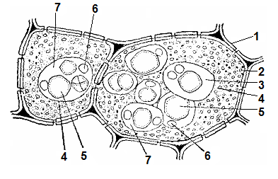
Draw the region of the seed transverse section containing complex grains in your laboratory notebook and label their constituent parts.
Conclusions. 1. A characteristic property of living cells is their ability to store carbohydrates, fats, and proteins as reserve nutrients.
2. Carbohydrates are stored as reserve substances, notably in the form of starch grains. Primary, transitory, and secondary (or reserve) starch are distinguished.
3. Proteins are stored as reserves in the form of aleurone grains, gluten, and crystals.
QUESTIONS FOR SELF-control
1. What compounds are stored as reserves in a plant cell?
2. Name the reserve nutrients.
3. Which reserve nutrients are formed in place of drying vacuoles?
4. What is meant by the terms oleoplasts and hydrophlasts?
5. Explain The process of starch grain formation in plants.
6. What is The Role of amyloplasts in The formation of reserve starch? How do they differ from other Plastids?
7. Name the types of starch grains found in various plant species.
8. Which organelles are involved in the formation of starch grains?
9. Name the oilseed crops cultivated in Ukraine.
Last update: 07/08/2026
Editorial and Educational Adaptation: This material has been compiled based on the primary/original source text. The project team performed an editorial review, corrected technical inaccuracies, structured sections, and adapted the content for an educational format.
What was processed:
- elimination of formatting defects (OCR errors, structural breaks, corrupted characters);
- editorial organization of content;
- standardization of terminology in accordance with academic sources;
- verification of factual statements against the original source text.
All mentions of the author, publication year, and origin of the primary text have been preserved in accordance with the source.