BIOCHEMISTRY - L. Stryer - 1984
VOLUME 3
PART V. MOLECULAR PHYSIOLOGY
CHAPTER 36. MEMBRANE TRANSPORT
36.9. Calcium transport is carried out by another ATPase
Calcium Ions play a crucial role in regulating Muscle contraction (Section 34.10), as well as many other physiological processes. Skeletal Muscle contains a complex network of membrane-bound tubules and vesicles. This membrane system, known as the sarcoplasmic reticulum, regulates the concentration of Ca2+ in the environment surrounding the contractile muscle fibers. At rest, Ca2+ is pumped into the sarcoplasmic reticulum, keeping the concentration of Ca2+ immediately surrounding the myofibrils extremely low. Excitation of the sarcoplasmic reticulum membrane by a Nerve Impulse triggers the instantaneous release of large amounts of Ca2+, which initiates muscle contraction. In other words, Ca2+ serves as the intermediate link between the nerve impulse and the contraction of the muscle fiber.
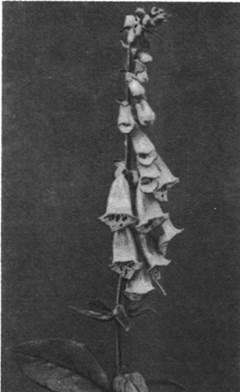
Class="center">Fig. 36.10. Foxglove
The transport of Ca2+ across the sarcoplasmic reticulum membrane is driven by ATP energy. The sarcoplasmic reticulum contains a Ca2+-activated ATPase. This Ca2+-ATPase constitutes the core of the Ca2+ pump, much like the (Na+ + K+)-ATPase forms the core of the (Na+ + K+)-pump. The Ca2+-ATPase also undergoes phosphorylation during ATP Hydrolysis:
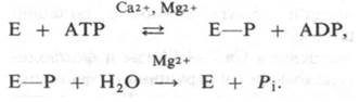
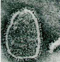
Fig. 36.11. Membrane vesicles formed from purified Ca2+-ATPase. The globular particles on the membrane surface represent portions of the ATPase molecule spanning the membrane
A cycle of conformational changes driven by phosphorylation and dephosphorylation mediates the translocation of two Ca2+ ions per cleaved ATP molecule. Due to the very high affinity of this ATPase for Ca2+ (K ~107 M), the enzyme efficiently transports Ca2+ from the Cytosol (where [Ca2+] <10-5 M) into the sarcoplasmic reticulum (where [Ca2+] ~ 10-2 M).
The density of Ca2+ pump molecules in the sarcoplasmic reticulum membrane is remarkably high, reaching approximately 20,000 per 1 μm2. In fact, the Ca2+-ATPase accounts for over 80% of the total integral Membrane Proteins and occupies a third of its surface area. The large subunit (100 kDa) of the Ca pump spans the membrane and contains the phosphorylation site; similarly to the (Na+ + K+)-pump, this site is a specific side chain provided by an aspartate residue. Another shared feature of both pumps is the presence of a glycoprotein: in the Ca2+ pump, a 55-kDa glycoprotein is associated with the large subunit.
A functionally active Ca2+ pump has been successfully reconstructed from purified Ca2+-ATPase and Phospholipids. In this Procedure, the Ca2+-ATPase was isolated from sarcoplasmic reticulum membranes following solubilization with the detergent cholate. The purified solubilized enzyme was added to soybean phospholipids. Upon removal of the detergent by dialysis, membrane vesicles were formed. These reconstituted vesicles rapidly accumulated Ca2+ in the presence of ATP and Mg2+.
36.10. The Na+ gradient powers the active transport of sugars and amino acids in animal cells
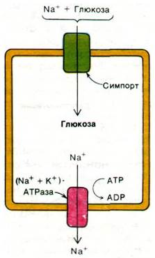
Many transport processes do not rely directly on ATP hydrolysis, but are instead coupled to the flow of ions down an electrochemical gradient. For instance, in many animal Cells, glucose accumulation is driven by the simultaneous influx of Na+. In this process, sodium ions and glucose bind to a specific transport protein and enter The Cell concurrently. This coordinated transfer of two components is termed cotransport; symport refers to the transport of both components in the same direction, whereas antiport involves opposite directions. The sodium ions that enter the cell alongside glucose molecules via symport are subsequently pumped out by the (Na+ + K+)-ATPase (Fig. 36.12). Both The amount of transported glucose and its transport rate depend on the transmembrane Na+ concentration gradient.
Fig. 36.12. The Na+ concentration gradient provides the energy for active glucose transport. This symport system is characteristic of the Plasma Membranes of intestinal and renal cells
Na+-dependent symport is widely utilized in animal cells for the accumulation of Amino Acids. In certain cells, such as those in the brush border microvilli of the intestine (Fig. 36.13), sugars are actively transported via symport mechanisms. Furthermore, the Small Intestine features a specialized Na+-dependent symport system that mediates the uphill transport of Cl- ions against their concentration gradient. In many cells, sodium ions also act as the driving force for antiport processes designed to extrude calcium ions. Thus, the sodium ion gradient established by the (Na+ + K+)-ATPase provides the energy for the majority of symports and antiports in animal cells.
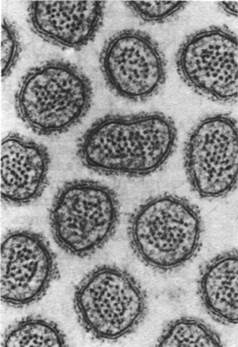
Fig. 36.13. Electron micrograph of a cross section through intestinal microvilli. The presence of microvilli vastly increases the surface area available for nutrient transport
36.11. Proton motive force drives numerous transport processes in bacteria
Symports and antiports are evolutionarily ancient mechanisms of molecular transport. For example, the flow of protons across The Plasma Membrane serves as the driving force for many transport systems in Bacteria. The best-characterized bacterial symport system is lactose transport in E. coli (Fig. 36.14). This resident of the lower mammalian intestine has evolved a highly efficient mechanism for concentrating lactose. Isolated by Eugene Kennedy, the pump for this disaccharide consists of a single 30-kDa polypeptide chain known as lactose permease (or the M protein). It is an integral membrane protein encoded by the y Gene, which is part of the lac Operon (Section 28.3). In induced cells, it accounts for approximately 4% of the membrane proteins.
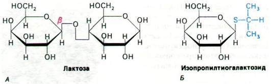
Fig. 36.14. Lactose permease transports β-galactosides, specifically lactose (A) and isopropyl thiogalactoside (B). Thiogalactosides have proven exceptionally useful for studying this system because they are transported by lactose permease yet escape hydrolysis by β-galactosidase
The Mechanism of the lactose pump was uncovered through The Study of y-gene mutants and investigations of vesicles derived from bacterial membranes. Vesicles are exceptionally useful for studying transport processes because their Structure is much simpler than that of an intact bacterium. While vesicles contain the Oxidative Phosphorylation system and other membrane-bound proteins, they lack the cytoplasmic components of an intact cell. Vesicles alone do not accumulate lactose; however, The addition of an oxidation substrate—which drives a flow of high-potential electrons along the Respiratory Chain—triggers lactose accumulation. The same effect can be achieved alternatively by establishing a pH gradient using extracellular acid. Generating a Membrane Potential via a K+ concentration gradient also drives lactose uptake. Taken together, these findings demonstrate that active lactose transport is powered by the proton-motive force across the plasma membrane. The transport of lactose molecules is coupled with the influx of a proton into the cell. Under physiological conditions, the proton gradient required for this Active Transport is generated by the flow of electrons from a high-potential donor (such as NADH) along the respiratory chain. This proton-lactose symport exemplifies Peter Mitchell's overarching concept of "energy conversion via a proton gradient" (Section 14.18).
Last update: 06/08/2026
Editorial and Educational Adaptation: This material has been compiled based on the primary/original source text. The project team performed an editorial review, corrected technical inaccuracies, structured sections, and adapted the content for an educational format.
What was processed:
- elimination of formatting defects (OCR errors, structural breaks, corrupted characters);
- editorial organization of content;
- standardization of terminology in accordance with academic sources;
- verification of factual statements against the original source text.
All mentions of the author, publication year, and origin of the primary text have been preserved in accordance with the source.