BIOCHEMISTRY - L. Stryer - 1984
VOLUME 1
PART I. CONFORMATION AND DYNAMICS
CHAPTER 8. ZYMOGEN ACTIVATION: DIGESTIVE ENZYMES AND COAGULATION FACTORS
The enzymatically active form of Lysozyme arises from the spontaneous folding of the molecule into the three-dimensional Structure characteristic of this enzyme. In contrast to lysozyme, many other Proteins are synthesized as inactive precursors that are subsequently activated by the Cleavage of one or more specific peptide bonds. While a catalytically active protein is called an enzyme, its inactive precursor is called a proenzyme (or zymogen).
Protein activation by specific proteolysis is a widespread process in biological systems. Here are a few Examples.
1. Digestive Enzymes that hydrolyze proteins are synthesized in the Stomach AND Pancreas as zymogens (Table 8.1).
Class="center">Table 8.1. Zymogens synthesized in The Stomach and pancreas

2. Blood clotting is mediated by a cascade of proteolytic activation reactions, providing a rapid and amplified response to injury.
3. Some protein Hormones are synthesized as inactive precursors. For example, Insulin is formed from proinsulin by the proteolytic removal of a peptide.
4. Collagen, a fibrous protein abundant in Skin and bone, is formed from a soluble precursor, procollagen.
8.1. Chymotrypsinogen is activated by the specific cleavage of a single peptide bond
Chymotrypsin is a digestive enzyme that hydrolyzes proteins in the Small Intestine. Like several other zymogens and digestive enzymes, it is synthesized in the pancreas as an inactive precursor, chymotrypsinogen. Indeed, the pancreas is one of the most active protein-synthesizing Organs. The enzymes and their precursors are synthesized in the acinar Cells of the pancreas (Fig. 8.1). Within these cells, Newly synthesized proteins are transported from The Endoplasmic reticulum to the Golgi apparatus, where they are enclosed in a protein-lipid membrane to form zymogen granules, which appear as highly dense bodies under an Electron microscope. The high electron density of zymogen granules is due to their high protein content (Fig. 8.2). Zymogen granules accumulate at the apex of acinar cells and are then secreted, in response to a hormonal or neural signal, into the duct leading to the duodenum.
Fig. 8.1. Schematic representation of zymogen secretion by a pancreatic acinar Cell
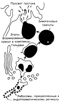
Fig. 8.2. Electron micrograph of zymogen granules in pancreatic acinar cells

Chymotrypsinogen consists of a single polypeptide chain of 245 Amino Acids cross-linked by five Disulfide Bonds. Chymotrypsinogen is virtually devoid of enzymatic activity. However, it is converted into an active enzyme when the peptide bond between Arginine-15 and isoleucine-16 is cleaved by Trypsin (Fig. 8.3). The resulting active enzyme, called П-chymotrypsin, then acts on other п-chymotrypsin molecules. The removal of two more Peptides yields the stable form of the enzyme, α-chymotrypsin. The subsequent cleavages in The conversion of п-chymotrypsin to the α-form are actually unnecessary, as л-chymotrypsin itself is fully active. The striking feature of this activation process is that the cleavage of a single specific peptide bond transforms the protein from a catalytically inactive form into a fully active one.
Fig. 8.3. Activation of chymotrypsinogen

Last update: 06/08/2026
Editorial and Educational Adaptation: This material has been compiled based on the primary/original source text. The project team performed an editorial review, corrected technical inaccuracies, structured sections, and adapted the content for an educational format.
What was processed:
- elimination of formatting defects (OCR errors, structural breaks, corrupted characters);
- editorial organization of content;
- standardization of terminology in accordance with academic sources;
- verification of factual statements against the original source text.
All mentions of the author, publication year, and origin of the primary text have been preserved in accordance with the source.