BIOCHEMISTRY - L. Stryer - 1984
VOLUME 1
PART I. CONFORMATION AND DYNAMICS
CHAPTER 8. ZYMOGEN ACTIVATION: DIGESTIVE ENZYMES AND COAGULATION FACTORS
8.2. Three-Dimensional Structure of Chymotrypsin
Understanding The Essence of this remarkable activation process requires a detailed knowledge of the Structure and catalytic Mechanism of Chymotrypsin. Fortunately, this enzyme has been extensively studied using chemical and X-ray crystallographic Methods. In fact, chymotrypsin is one of the most thoroughly investigated Enzymes, making a detailed examination highly worthwhile.
The chymotrypsin molecule consists of three polypeptide chains linked by two interchain Disulfide Bonds (Fig. 8.4). The molecular mass of the enzyme is approximately 25 kDa. Its three-dimensional structure at 2 Å resolution (Fig. 8.5) was determined by David Blow and coworkers using X-ray crystallography.
Class="center">Fig. 8.4. The α-chymotrypsin molecule contains two interchain disulfide bridges and three intrachain disulfide bonds

Fig. 8.5. Three-dimensional structure of α-chymotrypsin. Only α-carbon atoms are shown. Catalytic residues are highlighted in color
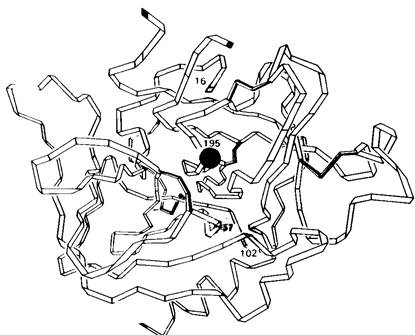
The chymotrypsin molecule is a compact ellipsoid measuring 51 x 40 x 40 Å. All charged groups, except for three that are absolutely essential for catalysis, are located On the surface of the molecule. The molecule is folded in a highly complex manner; unlike Myoglobin and Hemoglobin, it contains very little α-Helix. The polypeptide chains are mostly extended and often run in parallel strands spaced about 5 Å apart. Numerous Hydrogen Bonds form between peptide groups in adjacent strands. Part of the molecule possesses a Secondary structure resembling antiparallel β-pleated sheets, similar to what is observed in Lysozyme.
8.3. Chymotrypsin Is Specific for Aromatic and Large Nonpolar Side Chains
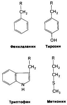
The Biological Role of chymotrypsin is to hydrolyze Proteins in the Small Intestine (Fig. 8.6). The equilibrium of this reaction is shifted almost completely toward Hydrolysis (> 99%). However, chymotrypsin does not rapidly hydrolyze just any peptide bond. It acts selectively on peptide bonds formed by the carboxyl groups of Amino Acids with aromatic side chains—Tyrosine, Tryptophan, and phenylalanine—as well as amino acids with large hydrophobic residues, such as Methionine (Fig. 8.7).
Fig. 8.6. Chymotrypsin catalyzes the hydrolysis of peptide and ester bonds

Chymotrypsin also hydrolyzes ester bonds. Although this reaction has no major physiological significance, it is of interest because it shares many features with peptide bond hydrolysis (Fig. 8.6). In fact, much of our knowledge about The Mechanism of chymotrypsin catalysis was obtained from studies of ester hydrolysis.
Fig. 8.7. Chymotrypsin preferentially hydrolyzes peptide bonds formed by the carboxyl groups of aromatic amino acids, as well as amino acids with large nonpolar side chains
8.4. During Chymotrypsin Catalysis, a Portion of the Substrate Is Covalently Bound to the Enzyme
Chymotrypsin catalyzes the hydrolysis of peptide and ester bonds in two distinct steps. This was first discovered during kinetic studies of p-nitrophenyl acetate hydrolysis. When using large amounts of the enzyme, two phases in the release of one of the reaction products, p-nitrophenol, can be clearly observed (Fig. 8.8). Initially, There is a rapid, burst-like release of p-nitrophenol, followed by its formation at a slower, steady-state rate.
Fig. 8.8. Two phases of p-nitrophenol formation can be observed after mixing chymotrypsin and p-nitrophenyl acetate
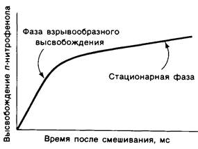
In the first step, p-nitrophenyl acetate binds to chymotrypsin, forming an enzyme-substrate (ES) complex (Fig. 8.9). The ester bond in the substrate is cleaved. One of the reaction products, p-nitrophenol, is released, while the acetyl group of the substrate becomes covalently bound to the enzyme. Next, Water attacks the acetyl-enzyme intermediate to yield an acetate ion and the regenerated enzyme (Fig. 8.10). The initial rapid phase of p-nitrophenol release corresponds to The formation of the acetyl-enzyme intermediate. This step is called acylation. The slower, steady-state formation of p-nitrophenol corresponds to the hydrolysis of the acetyl-enzyme intermediate and the regeneration of the free enzyme. This second step, called deacylation, is the rate-limiting step of the entire ester hydrolysis process by chymotrypsin. Furthermore, the acetyl-enzyme intermediate is stable enough to be isolated under favorable conditions. The mechanism of chymotrypsin catalysis can be represented by the following scheme:
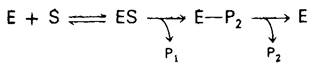
where P1 is the amine (or alcohol) component of the substrate, E-P2 is the intermediate formed via covalent bonding, and P2 is the acid component of the substrate.
Fig. 8.9. Acylation: Formation of the acetyl-enzyme complex as an intermediate

Fig. 8.10. Deacylation: hydrolysis of the intermediate acetyl-enzyme complex
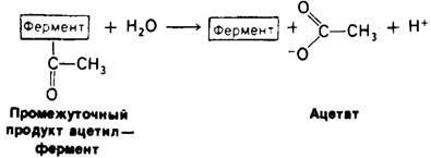
A distinctive feature of this reaction mechanism is the formation of an intermediate via covalent bonding; in the case discussed above, this involved the covalent attachment of an acetyl group to the enzyme. In general, the group attached to chymotrypsin at the E-P2 stage is always An acyl group. Thus, E2-P represents an acyl-enzyme intermediate.
Last update: 06/08/2026
Editorial and Educational Adaptation: This material has been compiled based on the primary/original source text. The project team performed an editorial review, corrected technical inaccuracies, structured sections, and adapted the content for an educational format.
What was processed:
- elimination of formatting defects (OCR errors, structural breaks, corrupted characters);
- editorial organization of content;
- standardization of terminology in accordance with academic sources;
- verification of factual statements against the original source text.
All mentions of the author, publication year, and origin of the primary text have been preserved in accordance with the source.