Pharmacognosy with the Basics of Plant Biochemistry - Kovalyov, V. M. 2004
Special Part
Phenolic Compounds
Anthraquinones and Other Anthracene Derivatives
Anthracene derivatives are compounds whose Structure is based on an anthracene Nucleus of varying degrees of oxidation, types of linkage, and monomeric Condensation.
Class="center">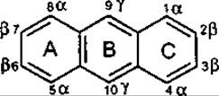
Anthracene
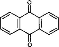
Anthraquinone
Reduced forms of anthraquinone—anthranols, anthrones, and oxanthrones—are easily oxidized to anthraquinone even by atmospheric oxygen under normal conditions, which is why anthraquinone derivatives are the most widespread and thoroughly studied.
They constitute the largest group of naturally occurring Quinones, with over 200 representatives currently known. Approximately half of these are found in plants. The most common anthraquinone is emodin, while the best-known anthraquinone in higher plants is alizarin—the primary pigment of common madder (Rubia tinctorum, Rubiaceae), which served as a major dye in ancient times.
Depending on The structure of the carbon Skeleton, anthracene derivatives are divided into two main groups: compounds based on a single anthracene derivative molecule (monomers) and compounds comprising two anthracene derivative molecules (dimers).
Monomeric anthracene derivatives. According to the degree of reduction of the anthraquinone core, this group is subdivided into two subgroups:
reduced forms—derivatives of anthranol, anthrone, and oxanthrone:
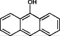
Anthranol
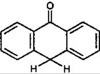
Anthrone
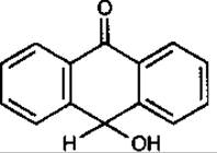
Oxanthrone
oxidized forms, which are based on the anthraquinone core.
Based on THE POSITION OF the OH groups in the molecule, monomeric anthraquinones are further divided into two subgroups:
emodin derivatives (chrysacin derivatives), or 1,8-dihydroxyanthraquinones, in which the OH groups are located on both benzene rings. These include emodin, chrysophanol, rhein, aloe-emodin, and others. These compounds and their derivatives act as laxatives;
alizarin derivatives, or 1,2-dihydroxyanthraquinones, in which the OH groups are located on a single benzene ring. These include alizarin, purpurin, lucidin, and others. These compounds and their derivatives exhibit nephrolithic activity.
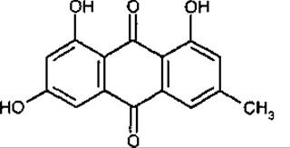
Emodin
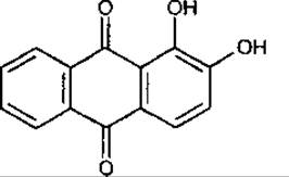
Alizarin
A special group of monomeric anthracene derivatives is represented by anthracyclines. Structurally, they feature a carbon skeleton in which an anthraquinone core is linearly fused to a six-membered saturated carbocycle, as seen in carminomycin.
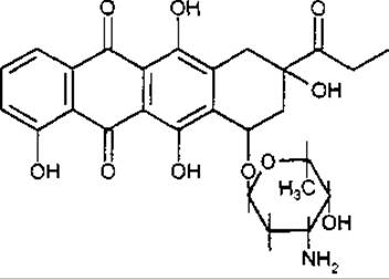
Carminomycin
Dimeric anthracene derivatives. Depending on the linkage type, dimeric anthracene derivatives are divided into linked by a single bond and condensed forms. Both reduced and oxidized forms occur in nature. The reduced dimers are typically linked via the у-position (chrysophanol dianthrone), whereas the oxidized forms are linked via the a- or ß-positions (casianin).
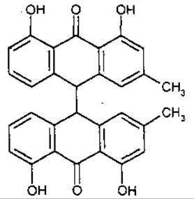
Chrysophanol dianthrone
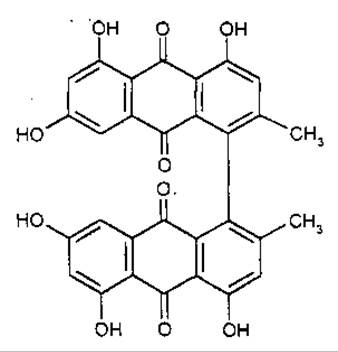
Casianin
Condensed anthracene derivatives differ from other dimeric compounds in that their monomeric skeletons are interconnected by two single bonds and one double bond, as exemplified by hypericin.
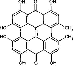
Hypericin
Most anthracene derivatives in natural sources occur as monomers in an oxidized form bearing various functional groups within the molecule: —ОН, —ОСН3, —СН2ОН, —СН3, —СОН, —СООН. Aryl substituents are extremely rare.
Anthracene derivatives occur either in a free state or as Glycosides. The aglycone moiety of anthraglycosides may belong to any group of anthracene derivatives, with the exception of dianthraquinones. The sugar component in glycosides is represented by glucose, rhamnose, xylose, arabinose, and biosides such as primverose, rutinose, and gentiobiose. The majority of anthraglycosides are O-glycosides. C-glycosides are significantly less common, occurring, for instance, in Aloe species.
Distribution and Localization
Anthracene derivatives are found in higher plants, Lichens, Fungi, Bacteria, insects, and marine animals of the phylum Echinodermata (crinoids). A substantial portion of anthraquinone derivatives has been isolated from fungi of the genera Aspergillus and Penicillium; in higher plants, anthraquinones are most frequently found in members of the families Rubiaceae, Rhamnaceae, Polygonaceae, Fabaceae, Asphodelaceae, Bignoniaceae, Verbenaceae, Scrophulariaceae, and others.
Reduced forms of hydroxyanthraquinones—anthranols, anthrones, and oxanthrones—are less commonly encountered in nature.
Anthracyclines are found in microorganisms, specifically streptomycetes (actinomycetes).
Anthracene derivatives accumulate in various plant parts, though most abundantly in leaves, fruits, bark, and underground Organs.
Anthracene derivatives are contained in a dissolved state within The Cell sap, and less frequently in dead plant Tissues.
Physicochemical Properties
Anthracene derivatives are crystalline substances ranging in color from yellow and orange to red. Free aglycones are soluble in ether, chloroform, benzene, and other organic Solvents, while being insoluble in Water.
Anthraglycosides are readily soluble in aqueous-alcoholic mixtures and water, less soluble in ethanol, and insoluble in benzene, chloroform, and ether. Both aglycones and glycosides readily dissolve in aqueous alkali solutions due to The formation of phenolates.
Hydroxymethylanthraquinones are yellow, orange, or red in color. Their coloration intensifies in alkaline solutions and concentrated sulfuric acid.
The hydroxyl group located at the a-position forms an intramolecular Hydrogen bond with the adjacent carbonyl group. This accounts for the differences in properties between the a- and ß-hydroxyl groups of the anthraquinone core. Hydroxymethylanthraquinones lacking an OH group in the ß-position are insoluble in carbonate and ammonia solutions, yet readily dissolve in alkali solutions; conversely, those possessing OH groups in the ß-position form salts with solutions of alkalis as well as carbonates and ammonia.
Hydroxymethylanthraquinones are stable toward high temperatures and oxidizing agents. For example, The oxidation of alizarin with manganese dioxide in sulfuric acid yields 1,2,4-trihydroxyanthraquinone.
Such oxidizing agents as nitric acid destroy anthraquinones, with rings lacking an OH group being oxidized to phthalic acids.
Similar to unsubstituted anthraquinone, hydroxyanthraquinones are reduced by sodium hydrosulfite in an alkaline medium to anthrahydroquinones.
Methods of Isolation and Analysis
Isolation. In crude plant Materials, free aglycones coexist with glycoside forms. If the goal is to obtain a mixture of anthracene derivatives for subsequent use without Separation, strong water-ethanol (70%) mixtures or pure alcohol (95%) are typically employed. When it is necessary to fractionate the total substance into individual components or fractions, fractional extraction is used. In cases where only aglycones are required, the glycosides undergo acid or enzymatic Cleavage, followed by the extraction of the total aglycones.
Selecting optimal conditions for separating the total anthraquinones into individual components is of critical importance. For this purpose, methods involving cleavage with salts and hydroxides of alkali and alkaline-earth metals are utilized. Thus, anthraquinones bearing a carboxyl group in their core dissolve in aqueous sodium bicarbonate and other alkaline solutions, whereas anthraquinones with a ß-hydroxy group do not form phenolates with sodium bicarbonate but react with solutions of alkali carbonates and hydroxides. If the anthraquinone core contains only a-hydroxyls, phenolates are formed exclusively in alkali solutions.
Nowadays, chromatographic methods are widely applied for separating all classes of compounds, including anthraquinones. Magnesium oxide, magnesol, silica gel, and ion-exchange resins are used for this purpose. Recently, polyamide resins have most frequently served as the sorbent.
Identification. Qualitative reactions and chromatographic methods are employed to detect anthracene derivatives.
The reaction with alkali solutions is the most specific, causing anthraquinones to turn red, while certain derivatives acquire a violet or black coloration.
According to the pharmacopoeial Procedure, the extraction of anthracene derivatives from plant materials is carried out using an alcoholic solution of potassium hydroxide under boiling conditions. After cooling, the filtrate is acidified with Hydrochloric acid until the red-brown color of the solution changes to yellowish-brown, and the acidic aqueous solution is extracted with diethyl ether. The ethereal extract is shaken with an ammonia solution; the ether layer remains yellow, while the alkaline layer turns red or violet.
Today, Thin-Layer Chromatography (TLC) and paper chromatography are the primary methods used to separate complex mixtures of natural compounds in order to identify specific classes of natural substances.
Following chromatographic separation, the plates or paper strips are treated with an alcoholic alkali solution. Spots of anthraquinone derivatives are identified by their yellow, red, or violet coloration.
Quantitative Assay. Nearly all Methods for the Quantitative determination of anthracene derivatives are based on assaying the total free hydroxymethylanthraquinones after prior Hydrolysis of anthraglycosides.
The photoelectrocolorimetric method proposed by Auterhoff is the most widely used. This method is included in the State Pharmacopoeia XI (SP XI) for assaying anthracene derivatives in Medicinal plant raw materials. The procedure involves extracting and hydrolyzing the anthracene derivative glycosides with glacial acetic acid, subsequently extracting them with an alkaline-ammonia solution, and measuring the optical density of the colored alkaline solution using a photoelectrocolorimeter. While the method is straightforward, result accuracy is affected by the reference standard used—cobalt chloride solution. A substance of anthraquinone nature, such as emodin or alizarin, should be employed as the standard reference.
Chromatospectrophotometric methods are used to determine individual compounds of anthraquinonic nature. Anthraquinones are extracted from medicinal plant materials and then separated via thin-layer or paper chromatography. The anthraquinone spots on the chromatograms are excised, the substance is extracted from them using an appropriate solvent, and the optical density is measured at the absorption maximum in the UV spectrum.
Biological Activity and Application
The biological activity of anthraquinones is highly diverse. They function as biochemical electron carriers in living organisms and participate in redox processes.
Anthraquinones of the emodin group enhance Large Intestine peristalsis, which accounts for their laxative effect. The laxative action manifests 10–12 hours after administration.
Alizarin derivatives from common madder (Rubia tinctorum) exhibit spasmolytic and diuretic effects and facilitate the elimination of renal calculi. Reduced forms of anthraquinone derivatives possess pronounced anti-inflammatory activity.
It has been established that condensed anthraquinones exhibit antitumor activity. A recent breakthrough has been the discovery of anthracyclines—Antibiotics possessing high antitumor efficacy.
Certain anthracycline derivatives exert inhibitory or stimulatory effects on enzymatic activity.
Information regarding plant materials and preparations containing anthraquinones is presented in Table 10 of the Appendix.
Last update: 06/08/2026
Editorial and Educational Adaptation: This material has been compiled based on the primary/original source text. The project team performed an editorial review, corrected technical inaccuracies, structured sections, and adapted the content for an educational format.
What was processed:
- elimination of formatting defects (OCR errors, structural breaks, corrupted characters);
- editorial organization of content;
- standardization of terminology in accordance with academic sources;
- verification of factual statements against the original source text.
All mentions of the author, publication year, and origin of the primary text have been preserved in accordance with the source.