Biochemistry - The Chemical Reactions of Living Cells, Volume 2 - D. Metzler 1980
Biosynthesis: How New Molecules Are Formed
Polyprenyl (Isoprenoid) Compounds
Carotenes and Their Derivatives
Although it has not yet been conclusively proven that carotenes are synthesized from phytoene, this is most likely the case. Fig. 12-14 illustrates the pathways for The formation of lycopene (the red pigment of tomatoes), ß-carotene, and other derivatives of these compounds. Note that The Structure of lycopene is entirely in the trans-configuration, and the desaturation process occurs via the loss of hydrogen atoms located in the trans-position. Desaturation proceeds in several stages, with many intermediates possessing a smaller number of double bonds having been identified. The ring closure at the ends of the lycopene molecule can be most easily conceptualized [Eq. (12-29)] via an acid-catalyzed mechanism involving a carbonium ion intermediate. The loss of either of the two protons adjacent to the positively charged carbon atom leads to the Formation of the ß-ring of ß-carotene or the a-ring of a-carotene, respectively [88].
Class="center">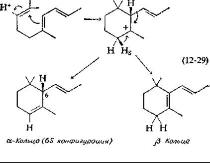
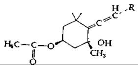
In the course of various reactions, carotenes can undergo Hydroxylation and other modifications. The structure of one of the resulting xanthophylls—zeaxanthin—is shown in Fig. 12-14. The reader will also find there the structure of fucoxanthin, the brown pigment of diatoms. Note that one end of the fucoxanthin molecule contains an epoxide group formed by the action of oxygen, while the other end bears a structure rarely encountered in nature: an allene group. (Quantitatively, fucoxanthin is likely the most abundant carotenoid [88].) Below we present the STRUCTURE OF THE allene-containing end of the fucoxanthin molecule (inverted compared to its depiction in Fig. 12-14). Please note that in Fig. 12-14 the Stereochemistry of the allene grouping is not depicted entirely correctly; specifically, the attached carotenoid chain R1 should project behind the ring, as shown below:
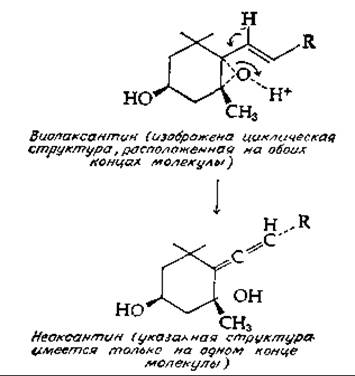
Another algal carotenoid, violaxanthin, contains epoxide groups in the cyclic structures at both ends of the molecule. In the presence of an isomerase, violaxanthin is converted [Eq. (12-30)] into neoxanthin, another compound presumably containing an allene structure (at one end). Subsequent Acetylation of this compound yields fucoxanthin.
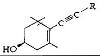
Some algal carotenoids contain acetylenic triple bonds. An example is alloxanthin, which bears the following symmetric structure at both ends of its molecule:
The description given above covers only a small fraction of the many structural modifications of carotenoids that have been described [44, 78, 89].
Supplement 12-B
Vitamins: Vitamin A
Vitamin A was discovered in the 1920s (Supplement 8-A); shortly thereafter, it was isolated from fish Liver oil. Both vitamin A1 (retinol) and vitamin A2 are 20-carbon polyprenol alcohols. They are formed by the Cleavage of 40-carbon ß-carotene (Fig. 12-14) or other carotenoids containing at least one ß-ionone ring. While carotenes are of plant origin, vitamin A is synthesized exclusively in animal Tissues. Under the action of an oxygenase, the carbon chain of carotene is centrally cleaved to yield vitamin A aldehyde, or retinalb. Vitamin A is present in tissues both as the free alcohol and as esters of palmitic and other Fatty acids. It is one of the few vitamins that can be stored in animal organisms in relatively large quantities. It is stored primarily as retinyl palmitate in specialized fat-storing Cells of the liverc.
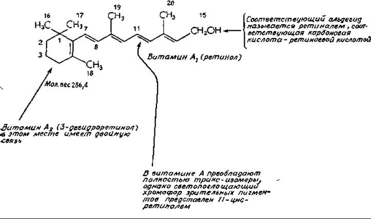
Vitamin A deficiency manifests with various symptoms, such as dry Skin and Hair, growth retardation, Conjunctivitis, and increased susceptibility to infection. A notable early sign of vitamin A deficiency is night blindness. Symptoms of Epithelial Tissue damage are most pronounced in the mucous membranes of the Lower Respiratory Tract and the Digestive System. The daily requirement of vitamin A for an adult human is approximately 0.7 mg. The vitamin A content in food is often expressed in International Units (IU): 1 mg of retinol corresponds to 3333 IU.
The participation of vitamin A in the form of retinal in visual function is well established (Chap. 13, Sec. E, 2); it also appears to play a specific role in reproduction. In vitamin A deficiency, males fail to produce spermatozoa, while females experience fetal resorption. Rats fed retinoic acid instead of vitamin A gradually become blind and sterile, but otherwise remain healthyd. Apparently, the alcohol or aldehyde form is required for reproductive function, whereas bone growth and the maintenance of normal secretory function of mucous membranes require only retinoic acide.
In vitamin A deficiency, the epithelium of Internal Organs—containing both specialized mucus-secreting cells and ciliated cells—becomes covered with a thick layer of keratinized, desquamating cells similar to those covering the external body surfaces. Concurrently, The production of fucose-containing glycopeptides drops sharplyf. It has been demonstrated that The addition of retinyl acetate to epidermal Cell cultures increases their RNA contentg. This effect becomes even more pronounced in the presence of Insulin and glucocorticoids. These findings indicate that retinoic acid or its metabolites likely serve as regulatory factors that drive the differentiation of unipotent epithelial cells into secretory mucosal cells. Another form of vitamin A presumably exerts a similar effect on reproductive tissues. In all likelihood, RNA METABOLISM/31.html">Transcription control is involved in mediating these regulatory Functions. Steroid Hormones and vitamin A derivatives apparently function in a similar manner, "turning on" or "turning off" genes that control the Synthesis of specific Proteins.
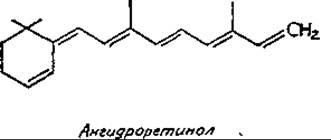
The chemical mechanism of vitamin A action remains unresolved. It can be hypothesized that retinal forms Schiff bases with protein amino groups, as occurs in visual pigments. The possible involvement of retinal in oxidation-reduction reactions cannot be excluded either. In the presence of HCl, retinol undergoes a non-enzymatic conversion to anhydroretinol via the elimination of a Water molecule, leaving behind a conjugated polyene system. Anhydroretinol occurs naturally, but apparently lacks biological activity:
In Blood Plasma, vitamin A is transported in a complex with a specific retinol-binding protein (MW ~21,000). This protein is normally almost fully saturated with retinol and is associated with another serum protein—prealbuminh,i. Retinol-binding proteins, as well as retinoic acid-binding proteins, are also present in many tissuesi. Evidence suggests that zinc is required to maintain normal blood plasma levels of vitamin Ak.
a Moore T., Vitamin A, Elsevier, Amsterdam, 1957.
b Olson J. A., Vitam. Horm. (N. Y.), 26, 1–63 (1968).
c Kobayashi K., Takahashi Y., Shibasaki S., Nature (London), New Biol., 243, 186–188 (1973).
d Smith J. E., Milch P. O., Muto Y., Goodman D. S., BJ, 132, 821–827 (1973).
e Clamon G. H., Sporn M. B., Smith J. M., Saffiotti V., Nature (London), 250, 64–66 (1974).
f DeLuca L., Schumacher M., Wolf G., JBC, 245, 4551–4558 (1970).
ж Sporn M. B., Dunlop N. M., Yuspa S. H., Science, 182, 722—723 (1973).
з Vahlquist A., Peterson P. A., Biochemistry, 11, 4526—4532 (1972).
и Muto Y., Smith J. E., Milch P. O., Goodman D. S., JBC, 247, 2542—2550 (1972).
к Ong D. E., Chytil F., JBC, 250, 6113—6117 (1975).
л Smith I. C., Jr., McDaniel E. G., Fan F. F., Halsted J. A., Science, 181, 954—955 (1973).
Last update: 06/08/2026
Editorial and Educational Adaptation: This material has been compiled based on the primary/original source text. The project team performed an editorial review, corrected technical inaccuracies, structured sections, and adapted the content for an educational format.
What was processed:
- elimination of formatting defects (OCR errors, structural breaks, corrupted characters);
- editorial organization of content;
- standardization of terminology in accordance with academic sources;
- verification of factual statements against the original source text.
All mentions of the author, publication year, and origin of the primary text have been preserved in accordance with the source.