Human Biochemistry Volume 2 - Murray R. 1993
Structure, Function, and Replication of Information Macromolecules
Recombinant DNA Technology
Basic Concepts of Genetic Engineering
The methodology of Introduction/32.html">Genetic Engineering is based on the Isolation of DNA and the performance of various manipulations with its molecules, including the creation of chimeras (for example, a DNA molecule constructed from human and bacterial DNA fragments).
Restriction Enzymes
One of the most essential tools in genetic engineering is endonucleases, which are enzymes that cleave DNA at specific nucleotide sequences within the chain (in contrast to exonucleases, which cleave DNA from the ends of the molecule). These enzymes are called restriction enzymes (or Restriction Endonucleases) because their presence in a bacterial Cell restricts the growth of certain bacterial Viruses known as Bacteriophages. Restriction enzymes cleave DNA into relatively small fragments at sequence sites with a strictly defined Structure. This mode of action distinguishes them from most other enzymatic, chemical, or physical treatments that cause random breaks in DNA strands. Restriction enzymes (over 200 types of enzymes in this Class have already been discovered) are part of the bacterial defense system that protects the host genome from foreign, primarily viral, DNA. Restriction enzymes are present only in Cells that also contain specific methylases. The Role of these enzymes is to methylate The Cell's own (host) DNA, thereby protecting it from the action of restriction enzymes. Site-specific methylases and restriction enzymes are always simultaneously present in Bacteria.

Fig. 36.1. Organization OF THE METABOLISM/31.html">Transcription unit and stages of EUKARYOTIC Gene Expression. Eukaryotic genes consist of structural and regulatory regions. The structural region is represented by coding DNA and non-coding 5'- and 3'-sequences. Coding DNA includes exons—DNA segments whose transcripts remain part of mature mRNA—and introns (transcripts of these sequences are removed from RNA during Processing). The structural region of a gene is bounded at the 5'-end by the Transcription initiation site and at the 3'-end by the polyadenylation or termination site. The promoter region, which contains specific sequences that interact with protein factors, is discussed in chapters 39 and 41. The primary transcript has a specific structure—a CAP—at the 5'-end and an "A"-rich region at the 3'-end. After the removal of introns from the primary transcript, the resulting mature mRNA is transported to the Cytoplasm, where it is translated to form a protein molecule.
Restriction enzymes are conventionally named after the bacteria from which they are isolated (Table 36.1). For instance, the name EcoRI indicates that this enzyme was isolated from Escherichia coli, and BamHI from Bacillus amyloliquefaciens. The first of the three letters in the abbreviation corresponds to the first letter of the genus name (E). The next two letters are the initial letters of the species name (co). The letter R indicates the specific strain from which the enzyme was isolated. The Roman numeral corresponds to the ordinal number of the restriction enzyme among similar enzymes isolated from that microorganism (e.g., EcoRI, EcoRII). Each enzyme recognizes a specific 4- to 7-base pair sequence in double-stranded DNA. Cleavage of DNA at these sites results in The formation of either "blunt" (e.g., produced by the restriction enzyme HpaI) or "sticky", i.e., overlapping (e.g., BamHI), ends (Fig. 36.2). Sticky ends are particularly useful for constructing hybrid molecules (see below). Assuming that NUCLEOTIDES are distributed along a DNA molecule in a completely random manner, the frequency of occurrence of the recognition site for a given restriction enzyme can be calculated. At any nucleotide position in a DNA molecule, any of the 4 nucleotides (A, G, C, T) has an equal probability of appearing. Therefore, an enzyme that recognizes a four-base sequence will find one target per 256 Base Pairs (44). At the same time, an enzyme that recognizes a six-base sequence will find a specific recognition site once every 4096 base pairs (46). Any DNA fragment has a characteristic arrangement of recognition sites for various restriction enzymes, making it possible to construct so-called restriction maps. Digestion of DNA with any single restriction enzyme yields a mixture of fragments, each sharing identical terminal segments. Such fragments can be separated by agarose or Polyacrylamide gel Electrophoresis (for blotting Methods, see below). This Procedure, based on The Use of restriction enzymes, represents a crucial step in Gene cloning.
Table 36.1. Selected restriction enzymes and their cleavage sequences

A — adenine; C — cytosine; G — guanine; T — thymine. Arrows indicate cleavage sites. Restriction can generate "sticky" (BamHI) or "blunt" (Hpa I) ends. The length of the recognized sequence varies and is 4 bp for Taq I, 5 bp for EcoRII, 6 bp for EcoRI, and 7 bp for Mst II. According to established conventions, the upper strand of the target sequence is presented in the 5'→3' orientation, and the lower strand in the 3'→5' orientation. Note that most sequences are palindromes (i.e., they read the same in opposite directions on different strands). The residue designated N represents any nucleotide.
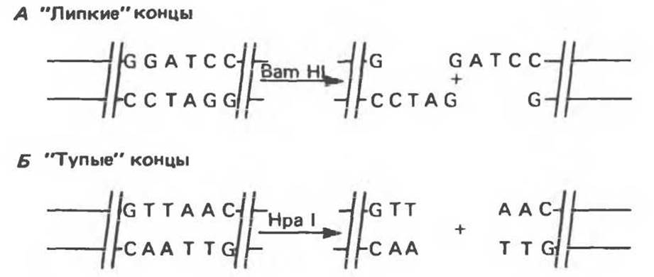
Fig. 36.2. Cleavage of DNA by restriction endonucleases (restriction enzymes). Restriction yields fragments (restriction fragments) with either "sticky" (A) or "blunt" (B) ends.
A number of other enzymes that use DNA AS A substrate are also vital for genetic engineering. Some of these will be discussed in this and subsequent chapters (Table 36.2).
Construction of Chimeric DNA Molecules
Covalent joining (hereinafter referred to as "ligation") of sticky ends of DNA fragments is technically straightforward, yet certain problems may arise. For instance, the sticky ends of a vector can ligate to themselves without incorporating the cloned fragment. In addition, annealing of sticky ends from two different fragments may occur, leading to the formation of a heterogeneous insert. Not all DNA segments of interest to researchers contain conveniently located recognition sites for restriction enzymes that produce sticky ends. To overcome these difficulties, restriction enzymes that generate blunt ends are sometimes used, followed by the generation of new ends using a specific enzyme (terminal transferase). For example, attaching a homopolymer chain consisting of dG to the 3'-ends of the vector and a poly-d(C) chain to the 3'-ends of the cloned DNA fragment ensures exclusively intermolecular annealing. During this procedure, known as "homopolymer tailing", a site for the restriction enzyme SmaI is also created, allowing for the subsequent excision of the cloned fragment. Occasionally, synthetic oligonucleotide linkers containing recognition sites for a specific restriction enzyme are attached to blunt-ended DNA. Direct ligation of blunt-ended fragments can also be performed using bacteriophage T4 DNA ligase. Although less efficient than sticky-end ligation, this method has the advantage of enabling the joining of any pairs of fragments. Its drawbacks include the inability to easily control the insert orientation and the number of inserted fragments, as well as the difficulty of "excising" the cloned fragment from the recombinant DNA molecule.
Table 36.2. Enzymes used in Recombinant DNA technology. (Adapted and reproduced, with permission, from Emery A. E. H. Page 41 in: An introduction to Recombinant DNA, Wiley, 1984.)
|
Enzyme |
Reaction |
Application |
|
Alkaline phosphatase |
Dephosphorylates the 5'-ends of RNA and DNA |
Removal of 5'-PO4 groups prior to end-labeling by kinasing, and Prevention of self-ligation of vector molecules |
|
BAL-31 nuclease |
Degrades both 5'- and 3'-ends of DNA |
Introduction of terminal deletions into DNA molecules |
|
DNA ligase |
Catalyzes the formation of phosphodiester bonds between DNA molecules |
"Joining" (ligation) of DNA molecules |
|
DNA polymerase I |
Synthesizes double-stranded DNA using a DNA template |
Synthesis of double-stranded cDNA; nick Translation |
|
DNase I |
Under appropriate conditions, introduces single-stranded breaks (nicks) into DNA |
Nick translation, mapping of hypersensitive sites in DNA |
|
Exonuclease III |
Removes nucleotides from the 3'-ends of DNA |
DNA Sequencing, mapping of protein-DNA interactions |
|
λ-Exonuclease |
Removes nucleotides from the 5'-ends of DNA |
DNA sequencing |
|
Polynucleotide kinase |
Transfers the terminal (γ-phosphate) group of ATP to 5'-OH groups of DNA and RNA |
Introduction of 32P into DNA and RNA |
|
Synthesizes DNA using an RNA template |
cDNA synthesis from mRNA; RNA mapping (from the 5'-end) |
|
|
S1 nuclease |
Degrades single-stranded DNA |
Removal of hairpin loops during cDNA synthesis; RNA mapping (from both 5'- and 3'-ends) |
|
Terminal transferase |
Adds nucleotides to the 3'-end of DNA |
Creation of homopolymer "tails" |
Cloning
A "clone" is defined as a large population of identical molecules, bacteria, or cells derived from a single common ancestor. Cloning makes it possible to obtain a vast number of identical DNA molecules that can be characterized and utilized for various purposes. The cloning method relies on the fact that chimeric or hybrid DNA molecules can be constructed within cloning vectors—such as bacterial Plasmids, phages, or cosmids—which are capable of Replication in host Cells under the control of their own regulatory elements. In this way, Amplification of the chimeric DNA is achieved. The general workflow of the cloning process is illustrated in Fig. 36.3.
Bacterial plasmids are small circular double-stranded DNA molecules whose Functions include, for example, conferring Antibiotic Resistance to the host cells carrying them. Plasmids possess several properties that make them exceptionally convenient as cloning vectors: 1) they can exist in a bacterial cell as single or multiple copies; 2) they replicate independently of the host DNA. Currently, the complete nucleotide sequence is known for many plasmids, which allows for the precise mapping of restriction sites for cloning DNA fragments. Plasmids are significantly smaller than the host chromosomal DNA and can therefore be easily separated from it. The cloned fragment is readily isolated from the recombinant plasmid by digesting it with the same restriction enzyme that was used for the initial ligation.
Phages typically contain linear DNA into which foreign DNA fragments can be inserted at any of the available restriction sites. Chimeric DNA is usually isolated after the recombinant phage completes its lytic cycle and mature infectious phage particles are released. The main advantage of phage vectors over Plasmid Vectors is that, whereas plasmids can typically accommodate DNA fragments up to 6–10 kb, phage particles can package fragments up to 10–20 kb in size. The size of the clonable fragment is determined by the total amount of DNA that can be packaged into the phage HEAD.
Even larger fragments can be cloned in cosmids—vectors that combine the advantages of both plasmids and phages. Cosmids are plasmids containing specific segments called cos sites, which are required for packaging phage λ DNA into the capsid. These vectors can be maintained within bacterial cells in plasmid form, but because most of the phage DNA has been removed from the cosmid, the allowable length of the cloned fragment is correspondingly increased. Inserts of 30–50 kb are quite common for cosmids. The properties of all three vector types are compared in Table 36.3.
When deciding into which region of a vector to introduce a cloned fragment, one must consider that an insertion into a functionally important sequence will inevitably disrupt one of the vector's properties. However, this disruption can be exploited for the Selection of recombinant molecules. For example, the widely used plasmid vector pBR322 carries genes for resistance to two Antibiotics—ampicillin and tetracycline. Cloning frequently utilizes a unique PstI site within the ampicillin resistance gene. The insertion of a foreign DNA fragment leads to the inactivation of the ampicillin resistance gene, and bacteria carrying such a recombinant plasmid become sensitive to this antibiotic (Fig. 36.4). This makes it easy to distinguish the original plasmid, which confers resistance to both antibiotics in the host bacterium, from the recombinant form. To obtain even more conclusive evidence that the plasmid DNA is indeed chimeric (carries an insert), the size of the constructed plasmid can be compared with that of the original plasmid using gel electrophoresis. Naturally, the recombinant plasmid will have a higher molecular weight.

Fig. 36.3. Use of restriction enzymes to construct recombinant or chimeric DNA molecules. When introduced back into a bacterial cell via transformation, the plasmid replicates, and along with it, the inserted sequence replicates as well. Because the reunion of sticky ends recreates the restriction enzyme recognition site, the cloned fragment can be easily excised from the recombinant plasmid using the same restriction enzyme. If a mixture of all fragments generated by digesting total human DNA with the same endonuclease is used, cloning plasmids can yield approximately one million different recombinant DNA molecules, each giving rise to an individual bacterial clone. (Modified and reproduced with permission, from Cohen SN: The manipulation of genes. Sci. Am. [July] 1975; 233: 34.)
Table 36.3. Common cloning vectors
|
Vector |
Size of cloned DNA fragment (insert) |
|
Plasmid pBR322 |
0.01—10 kb |
|
Lambda Charon 4A |
10—20 kb |
|
Cosmids |
35—50 kb |
Genomic Libraries and their construction
By selecting appropriate cloning conditions, it is possible to ensure that a set of cloned fragments contains virtually all The genes of a given genome. Such collections of clones obtained for a specific genome are called genomic libraries. A genomic library is prepared from total cellular DNA of a cell line or tissue. Unlike a genomic library, a cDNA library represents the mRNA population of a tissue. A genomic library is prepared by partial digestion of total DNA with a "frequent-cutting" restriction enzyme (e.g., SauIIIA). The purpose of this Treatment is to obtain a population of large DNA fragments containing full-length genes. Phage vectors are preferred for constructing genomic libraries because they allow the cloning of very long DNA fragments (up to 20 kb). The number of independently obtained phage vectors required to create a representative library is inversely proportional to the size of the cloned fragments, but directly proportional to the Genome Size (Table 36.4). A Human Genome library consisting of 106 clones with sufficiently large inserts contains any unique gene with a probability of about 99%. Obtaining such a library ensures a high probability of identifying the gene of interest.

Fig. 36.4. Method for identifying recombinant DNA molecules. A DNA fragment is inserted into the unique Pst I site of plasmid pBR322. The "insert" inactivates the gene encoding the protein that confers resistance to ampicillin in the host bacterium. Consequently, bacterial cells carrying the recombinant DNA are unable to form colonies on media containing this antibiotic. By analyzing transformants on media with tetracycline and ampicillin, clones carrying recombinant plasmids can be selected.
A cDNA library is prepared in several steps. First, total tissue mRNA is isolated, and then reverse transcription of mRNA into double-stranded DNA is performed using reverse transcriptase and DNA polymerase. For technical reasons, it is rarely possible to obtain a full-length copy of mRNA (cDNA). As a rule, shorter fragments are cloned. Plasmids are usually used for this work because they are easier to handle than phage and cosmid vectors. However, There is a whole class of vectors—lambda phages—specifically designed for cDNA cloning (see below).
Vectors that ensure the Synthesis of the protein encoded by the cloned gene are called expression vectors. Such vectors are widely used to identify specific cDNA molecules in a cDNA library, as well as to produce engineered Proteins. Specially engineered expression vectors typically possess a strong inducible promoter, Translation initiation codons for all possible reading frames, transcription and translation terminators, and, if necessary, specific protein processing signals. Some expression vectors contain protease inhibitor genes, which increase the yield of the expressed gene product. The λgt 11 vector is very frequently used for constructing cDNA libraries. This vector allows the cloning of sufficiently long cDNA fragments while ensuring the transcription and Translation of the cloned genes. Both cDNA probes and specific Antibodies are suitable for screening cDNA libraries based on the λgt 11 vector.
Table 36.4. Composition of representative genomic libraries 11
|
Source |
Representative genomic library |
|
E. coli |
1500 fragments |
|
4500 fragments |
|
|
Drosophila |
50,000 fragments |
|
Mammals |
800,000 fragments |
1) The number of fragments (independent clones) required to create a library guaranteed to represent all unique genes is inversely proportional to the average size of the cloned fragments and directly proportional to the total number of genes in the Organism. The numbers given above are for libraries with a 99% probability of representing the complete genome at a cloned fragment size of 2∙104 base pairs. Differences in the required number of individual clones reflect the varying degrees of genome complexity of the respective organisms.
The required number of clones is calculated using the formula

where P is the desired probability, and f is the fraction of the total genome contained within an individual clone. For mammals, characterized by a haploid genome complexity of 3∙109, this equation takes the following form:

The advantage of libraries constructed using large inserts becomes apparent when performing the corresponding calculations for a hypothetical insert with an average size of 5∙103 instead of 2∙104.
Probes
Various types of molecular probes are used to identify a specific clone within genomic or cDNA libraries, as well as for the Quantitative determination of DNA or RNA. Typically, probes are DNA or RNA fragments containing radiolabeled 32P nucleotides. The use of probes is based on their ability to "recognize" complementary sequences in DNA or RNA molecules. cDNA synthesized on a specific mRNA template can be used as a probe when screening cDNA and genomic libraries. One of the most common methods for searching for specific sequences is based on the use of synthetic oligonucleotides, whose nucleotide sequences are designed based on the Amino Acid Sequence of a small region of the target protein, taking into account codon degeneracy. Given an exact sequence match, an oligonucleotide probe length of 15–20 residues is sufficient for reliable Hybridization and detection of a unique gene. cDNA probes are also used to detect specific DNA or RNA fragments after electrophoresis and transfer to a nitrocellulose filter (Southern blotting and Northern blotting methods, respectively).
Blotting and Hybridization Techniques
To visualize a specific DNA or RNA fragment among thousands of other contaminant molecules, a combination of several experimental Procedures is used, collectively known as "blotting." Fig. 36.5 illustrates the schemes for Southern blotting (for DNA fragment visualization), Northern blotting (for RNA), and Western blotting (for proteins). The first procedure is named after the inventor of the method, while the others originated as laboratory jargon but eventually became generally accepted terms. The first technique is useful for determining gene copy number in a given tissue or for identifying significant structural alterations in genes (deletions, insertions, or rearrangements). Sometimes it is even possible to detect a point mutation if it affects a restriction site. Northern and Western blotting variants are used to determine the molecular sizes and quantities of specific RNA and Protein molecules.
To identify and isolate clones of interest to researchers, colony hybridization and plaque hybridization methods have been developed. Nitrocellulose filters are first applied to bacterial colonies grown on solid media. Bacteria adhere to the filter. Following lysis and Denaturation with NaOH and immobilization of the denatured DNA by baking, the filter is incubated in a solution containing a radiolabeled probe. After hybridization is complete, the filter is washed to remove excess probe, and the resulting labeled hybrid complex is detected by exposure to X-ray film. By comparing THE POSITION OF the spot on the autoradiogram with the position of the colonies on the plate, the colony that gave a positive signal is selected. A similar approach is used to identify clones based on phage vectors. The result of these manipulations is the Isolation of the desired individual clones (bacterial colonies or phage plaques).
All variants of hybridization methods discussed in this chapter are based on the aforementioned specific base-pairing interactions of complementary nucleic acid strands. An exact sequence match between hybridizing fragments leads to the rapid formation of a stable complex resistant to high temperatures during hybridization and washing. Such complexes are also stable at low salt concentrations. Complexes formed with relatively weaker sequence complementarity are less stable under stringent conditions (high Temperature or low salt concentration). In this case, either hybridization does not occur at all, or the hybrid complex is disrupted during washing. Gene families exhibiting some degree of Homology can be identified by varying hybridization and washing stringency. This same approach is applied when comparing homologous Genes from different species.
DNA Sequencing
Methods for determining the complete nucleotide sequence of DNA have now been developed (Fig. 36.6). Solving this problem requires a large quantity of identical DNA molecules. The sequence of interest can be amplified by cloning the corresponding fragment. The Maxam-Gilbert sequencing method, illustrated in Fig. 36.6, is based on the chemical cleavage of DNA at specific bases. Another enzymatic method—the Sanger method—is based on the use of nucleotide analogs that terminate the synthesis of the complementary DNA strand on a single-stranded template at the site of analog incorporation into the chain.

Fig. 36.5. Blotting transfer. In Southern blotting, total DNA isolated from a cell culture or tissue is treated with one or more restriction enzymes, and the resulting mixture of fragments is subjected to electrophoresis in an agarose or polyacrylamide gel. Negatively charged DNA migrates toward the anode, with smaller fragments moving faster than larger ones. Following electrophoresis, the separated DNA fragments undergo mild denaturation by incubating the gel in an alkaline solution. In the next step, the gel is placed onto a nitrocellulose filter. The DNA fragments are transferred to the nitrocellulose using methodological procedures developed by Southern, and the resulting nitrocellulose replica is fixed by heat treatment. Next, the replica is incubated with a labeled cDNA probe that hybridizes with the corresponding complementary DNA fragment on the nitrocellulose filter. After intensive washing, the filter is placed on an X-ray film. The signals captured on the autoradiograph correspond to the positions of the DNA fragments complementary to the probe sequence. The Northern blot method (for RNA analysis) is fundamentally similar to the Southern transfer procedure (Southern blot analysis). Total RNA is subjected to electrophoresis. The actual procedure for transferring RNA from the gel to the filter differs somewhat from the Southern method because RNA molecules are less stable than DNA molecules. The Western blot method is used to detect specific proteins using specific antibodies or other molecular probes.
Last update: 06/08/2026
Editorial and Educational Adaptation: This material has been compiled based on the primary/original source text. The project team performed an editorial review, corrected technical inaccuracies, structured sections, and adapted the content for an educational format.
What was processed:
- elimination of formatting defects (OCR errors, structural breaks, corrupted characters);
- editorial organization of content;
- standardization of terminology in accordance with academic sources;
- verification of factual statements against the original source text.
All mentions of the author, publication year, and origin of the primary text have been preserved in accordance with the source.