Human Biochemistry, Volume 1 - Murray R. 1993
Bioenergetics and Carbohydrate and Lipid Metabolism
Physiologically Important Carbohydrates
Polysaccharides
Polysaccharides include the following Physiologically Important CARBOHYDRATES.
Starch. In starch, monosaccharide residues are linked by a-glucosidic bonds. A Structure of this type, formed exclusively by glucose residues, is a homopolymer known as a glucosan or glucan. It is the most important dietary carbohydrate, found in cereals, potatoes, legumes, and other plants. The two Main Components of starch are amylose (15–20%), which has an unbranched helical structure (Fig. 14.14), and amylopectin (80–85%), consisting of branched chains where each branch comprises 24–30 glucose residues joined by (1→4)-bonds [at branching points, the residues are linked by (1→6)-bonds].
Class="center">
Fig. 14.13. Structure of several important Disaccharides; the a- and ß-forms differ in configuration at the anomeric carbon atom (marked with an asterisk). If the anomeric carbon of the second sugar residue participates in the glycoside bond, this residue is termed a glycoside (furanoside or pyranoside).
Table 14.3. Disaccharides
|
Sugar |
Source |
Clinical Significance |
|
Maltose |
Breakdown product of starch (via amylase Digestion or Hydrolysis). Germinating cereals, malt |
|
|
Lactose |
Milk. May be present in urine during Pregnancy |
Decreased lactase activity impairs lactose digestion, leading to diarrhea and flatulence |
|
Sucrose |
Cane and beet sugar. Sorghum, pineapple, carrot |
Decreased sucrase activity impairs sucrose digestion, resulting in diarrhea and flatulence |
|
Trehalose |
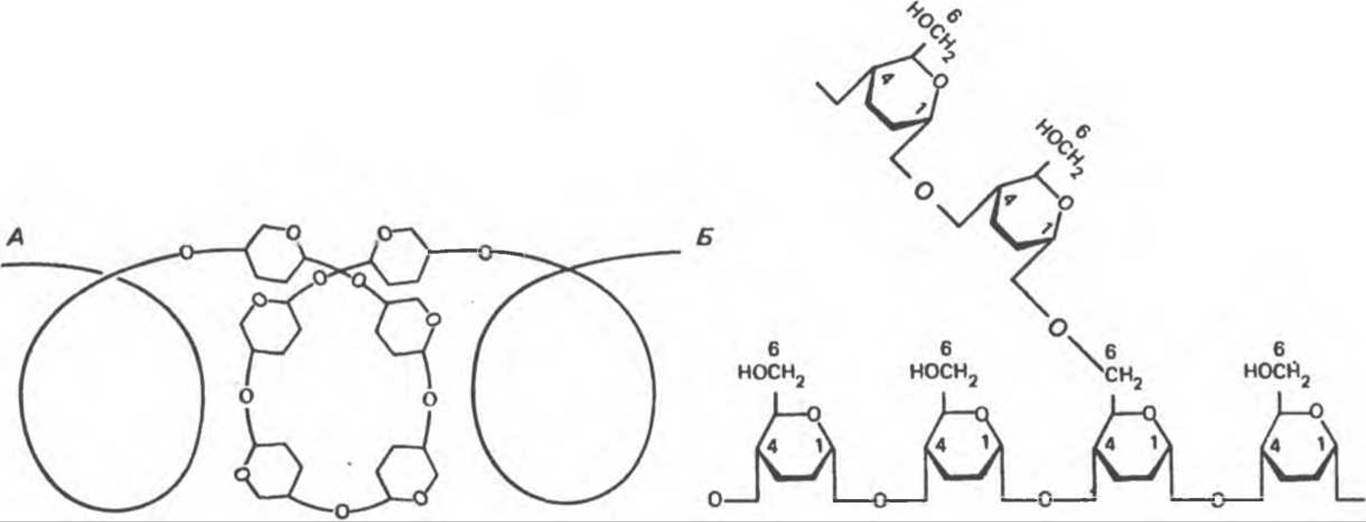
Fig. 14.14. Structure of starch. A — amylose with its characteristic helical structure; B — amylopectin, forming 1→6 type bonds at the branch points.
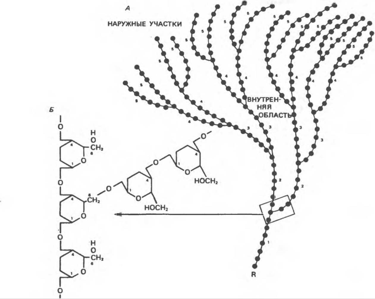
Fig. 14.15. Glycogen molecule. A — enlarged view of the structure near a branch point. B — molecular structure. Numbers indicate regions formed at equivalent stages of macromolecular growth. R — first glucose residue. Typically, branching is more complex than illustrated; The ratio of 1→4 bonds to 1→6 bonds ranges from 12 to 18.
Glycogen (Fig. 14.15) is the polysaccharide used to store carbohydrates in animal Tissues. It is frequently referred to as animal starch. Glycogen features a more highly branched structure than amylopectin, with linear segments containing 11–18 a-D-glucopyranose residues [joined by a(1→4)-glycosidic bonds], while at the branch points the residues are linked by a(1→6)-glycosidic bonds.
Inulin is a polysaccharide found in the tubers and roots of dahlias, artichokes, and dandelions. Upon hydrolysis, it yields fructose, making it a fructosan. Unlike potato starch, this polysaccharide is readily soluble in warm Water and is used in physiological research to determine the Glomerular Filtration rate in Kidneys.
Dextrins are substances formed during the Hydrolysis of Starch. The products formed at a specific stage of hydrolysis are termed "limit dextrins."
Cellulose is the primary structural component of plants. It is insoluble in common Solvents and consists of a-D-glucopyranose units linked by ß(1→4)-bonds, forming long, extended chains stabilized by cross-linking Hydrogen Bonds. Many mammals, including humans, cannot digest cellulose because their digestive tracts lack Hydrolases capable of cleaving ß-bonds. Therefore, cellulose can be considered a substantial unused dietary reserve. The gut of ruminants and other herbivores contains microorganisms capable of enzymatically breaking down ß-bonds, making cellulose an important source of dietary calories for these animals.
Chitin is an essential structural polysaccharide in invertebrates, forming, among other things, the exoskeleton of crustaceans and insects. The structure of chitin is composed of N-acetyl-D-glucosamine units linked by ß(1→4)-glycosidic bonds (Fig. 14.16).
Glycosaminoglycans (mucopolysaccharides) consist of complex carbohydrate chains containing amino sugars and uronic acids. When these chains are attached to a protein molecule, the resulting compound is called a proteoglycan. As a primary cementing substance, glycosaminoglycans are associated with Structural components of bone, as well as with Elastin and Collagen. Their function is to bind large amounts of water and fill intercellular spaces. They serve as cushioning and lubricating agents for various tissue structures; these Functions are facilitated by a high density of —OH groups and negative charges on their molecules, which cause mutual repulsion of the carbohydrate chains and prevent them from aggregating. Examples include hyaluronic acid, chondroitin sulfate, and heparin (Fig. 14.16), which will be discussed in detail in Chapter 54.
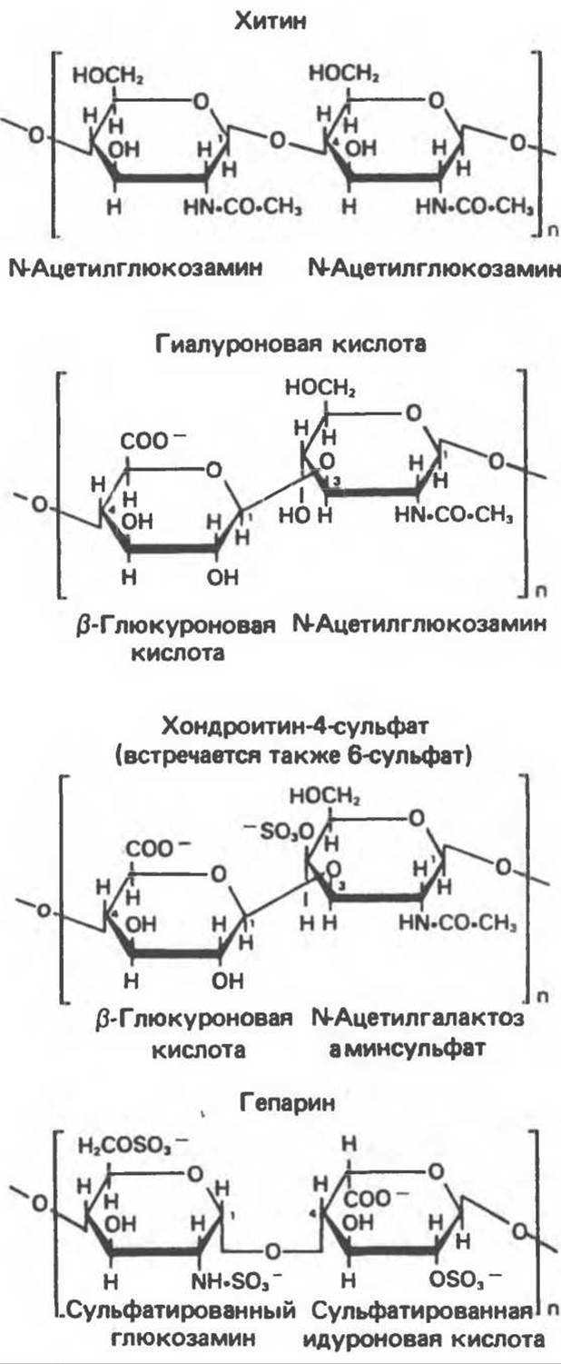
Fig. 14.16. Structure of some complex polysaccharides
Glycoproteins (mucoproteins) are found in various Body Fluids and tissues, as well as in Cell membranes (see Chapters 42 and 54). They are complex Proteins containing a carbohydrate component (in varying amounts) that may consist of short or long (up to 15 units), branched or unbranched chains. These chains, commonly referred to as oligosaccharide chains, include
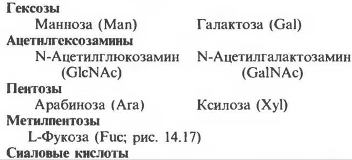
N-acyl derivatives of neuraminic acid, such as N-acetylneuraminic acid (NeuAc; Fig. 14.18), which is the predominant sialic acid.
Glucose is absent in fully formed (mature) glycoproteins (with the exception of collagen). Furthermore, unlike Glycosaminoglycans and Proteoglycans, glycoproteins do not contain uronic acids.
Sialic acids are N- or O-acyl derivatives of neuraminic acid (Fig. 14.18). Neuraminic acid is a nine-carbon sugar derived from mannosamine (an epimer of glucosamine) and Pyruvate. Sialic acids are constituents of glycoproteins and gangliosides.
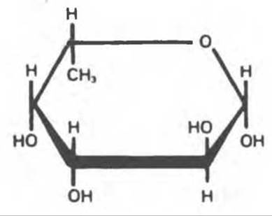
Fig. 14.17. ß-L-Fucose (6-deoxy-ß-L-galactose).

Fig. 14.18. Structure of N-acetylneuraminic acid, one of the sialic acids (Ac = CH3—CO—).
Last update: 06/08/2026
Editorial and Educational Adaptation: This material has been compiled based on the primary/original source text. The project team performed an editorial review, corrected technical inaccuracies, structured sections, and adapted the content for an educational format.
What was processed:
- elimination of formatting defects (OCR errors, structural breaks, corrupted characters);
- editorial organization of content;
- standardization of terminology in accordance with academic sources;
- verification of factual statements against the original source text.
All mentions of the author, publication year, and origin of the primary text have been preserved in accordance with the source.