Principles of Biochemistry Volume 1 - A. Lehninger 1985
Biomolecules
Water
Phosphate and bicarbonate are important biological buffer systems
In All living organisms, intracellular and extracellular fluids typically maintain a characteristic and constant pH value, which is regulated by various biological systems. However, the primary line of defense protecting living systems from internal pH fluctuations is provided by buffer systems. Two of the most
important buffer systems in mammals are the phosphate and bicarbonate systems. The phosphate buffer system, which plays a vital role in maintaining intracellular pH, consists of a conjugate acid-base pair formed by the H2PO-4 ion (a proton donor) and the HPO4 ion (a proton acceptor).
Class="center">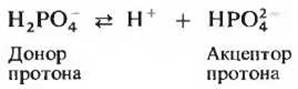
The phosphate buffer system operates in much the same way as the acetate system, except that it Functions within a different pH range. This system achieves maximum efficiency near pH 6.86 because the pK' value of H2PO-4 ions is 6.86 (see Table 4–4 and Fig. 4–11). The H2PO-4 — HPO2-4 phosphate buffer pair can resist pH changes between 6.1 and 7.7, thereby providing adequate buffering capacity for intracellular fluid, whose pH typically ranges from 6.9 to 7.4.
The principal buffer system of Blood Plasma is the bicarbonate system, which is a conjugate acid-base pair consisting of carbonic acid (H2CO3) as the proton donor and the bicarbonate ion (HCO-3) as the proton acceptor:

This system, which has its own Equilibrium Constant
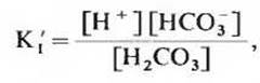
functions as a buffer in the same manner as other conjugate acid-Base Pairs. Its unique feature, however, is that one of its components—namely, carbonic acid (H2CO3)—is formed by the reaction of dissolved carbon dioxide (p) with Water in a reversible process:

the equilibrium constant of which is
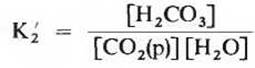
Since Carbon dioxide is a gas under normal conditions, the value of [СО2(р)], i.e., the concentration of dissolved СО2, is determined by equilibrium with СО2 in the gas phase (g):

characterized by the equilibrium constant K3, which is equal to
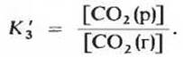
The pH of the bicarbonate buffer system depends on the concentrations of its dissolved components, H2CO3 and HCO-3, which act as proton donor and acceptor. However, because the concentration of H2CO3 depends in turn on the concentration of dissolved СО2—and the latter on the partial pressure of СО2 in the gas phase—the pH of a bicarbonate buffer in contact with a gas phase is ultimately determined by the concentration of HCO3 ions in the aqueous phase and the partial pressure of СО2 in the gas phase (see Box 4–3).
The bicarbonate buffer system functions as an efficient physiological buffer near pH 7.4 because the proton donor H2CO3 in blood plasma is in mobile equilibrium with a large reserve pool of gaseous СО2 in the air space of the Lungs. Whenever blood must absorb excess OH- ions for any reason and the pH rises, The amount of carbonic acid (H2CO3) partially converted to HCO3 through interaction with OH- ions is rapidly replenished from the large reservoir of gaseous СО2 in the lungs.
Box 4–3. How the Blood Bicarbonate System Works
The blood buffer system involves three interrelated reversible equilibria between gaseous СО2 in the lungs and the bicarbonate ion (HCO-3) in blood plasma (Fig. 1). When H+ ions enter the blood as it flows through tissue vessels, their concentration rises immediately. This causes the equilibrium of reaction 3 (Fig. 1) to shift, establishing a new equilibrium corresponding to a higher concentration of H2CO3, which in turn leads to an increased concentration of СО2 (р) in the blood. As a result, the СО2 pressure in the lung gas phase also rises, and the excess СО2 is exhaled. Conversely, when a certain amount of OH- ions enters the blood plasma, the events proceed in reverse. A decrease in H+ ion concentration causes some H2CO3 molecules to dissociate into H+ and HCO-3 ions, which in turn prompts the dissolution of additional СО2 (g) from the lungs into the blood plasma. Thus, a high rate of Respiration—that is, a high rate of inhaling air and exhaling СО2—can drive these equilibria rapidly enough to maintain a constant blood pH.

Fig. 1. An equilibrium is established between СО2 in the air space of the lungs and the bicarbonate buffer in blood plasma flowing through the pulmonary capillaries. Because the concentration of dissolved СО2 can be rapidly adjusted by altering the respiration rate, the blood bicarbonate buffer system remains nearly in equilibrium with an extensive potential reservoir of СО2.
СО2 (g) dissolves in the blood to form СО2 (р), which reacts with water to yield H2CO3 (see Box 4–3). Conversely, whenever blood pH decreases for any reason, some HCO3 of the buffer system binds with excess H+ ions, producing an excess of H2CO3. This H2CO3 breaks down, releasing dissolved СО2, which then passes into the gas phase in the lungs and is ultimately exhaled by the Organism. As blood flows through the numerous capillary vessels in the lungs, its bicarbonate buffer system rapidly approaches equilibrium with СО2 in the lung gas space. The combined operation of the bicarbonate buffer system and the lungs represents a crucial mechanism for maintaining a constant blood pH.
Blood plasma pH is maintained at a remarkably constant level. Under normal conditions, blood plasma has a pH close to 7.40. Disruptions in the mechanisms regulating pH—such as those observed in severe diabetes due to acidosis caused by the overproduction of metabolic acids—cause blood drop to pH 6.8 and below, which in turn can lead to irreversible consequences and death. In certain other pathological conditions, blood pH occasionally reaches values so high that normalization becomes impossible. Since an increase in H+ ion concentration of just 1.18×10⁻⁷ M (the approximate difference between blood at pH 7.4 and blood at pH 6.8) can be life-threatening, the question arises: what molecular mechanisms ensure that cellular pH is maintained with such high precision? Although pH influences many Structural and functional Properties of the Cell, the catalytic activity of Enzymes is particularly sensitive to pH changes. Figure 4–13 shows typical curves illustrating the dependence of various enzyme activities on pH. As can be seen, each of these enzymes exhibits maximum activity at a specific pH value known as the pH optimum. Deviation of pH in either direction from this optimal value is frequently accompanied by a sharp drop in enzymatic activity. Thus, minor shifts in pH can lead to substantial Changes in the rates of vital enzymatic reactions occurring, for instance, in Skeletal Muscle or the Brain. Biological control ensuring constant pH in Cells and Body Fluids is therefore of paramount importance for all aspects of METABOLISM and cellular activity.
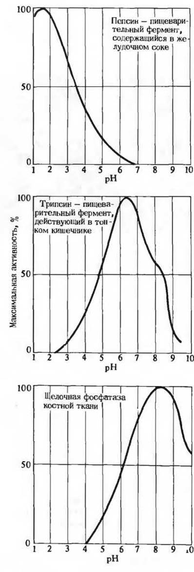
Fig. 4-13. Effect of pH on The activity of some enzymes. Each enzyme has a characteristic pH-activity profile.
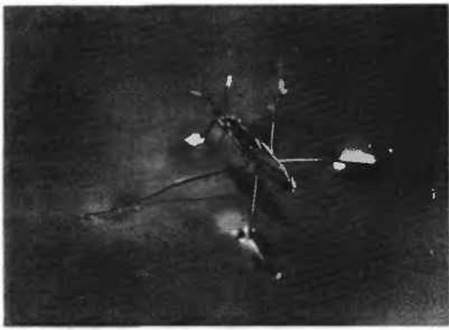
Fig. 4-14. A water strider (family Gerridae) taking advantage of the high surface tension of water. This pond-dwelling insect has specialized hairs on its first and third pairs of legs that allow it to stay atop the surface film without breaking it. The middle pair of legs penetrates the surface layer and acts as oars.
Last update: 06/08/2026
Editorial and Educational Adaptation: This material has been compiled based on the primary/original source text. The project team performed an editorial review, corrected technical inaccuracies, structured sections, and adapted the content for an educational format.
What was processed:
- elimination of formatting defects (OCR errors, structural breaks, corrupted characters);
- editorial organization of content;
- standardization of terminology in accordance with academic sources;
- verification of factual statements against the original source text.
All mentions of the author, publication year, and origin of the primary text have been preserved in accordance with the source.