INVERTEBRATE ZOOLOGY IN THREE VOLUMES - VOLUME 3 - H.I. Shcherbak - 1997
PHYLUM BRACHIOPODA
CLASS BRACHIOPODA
Brachiopods are exclusively marine animals inhabiting waters of normal salinity. Their sizes are mostly within the centimeter range, with the largest living species, Magellania venosa, reaching 8.4 cm. The entire soft body of a brachiopod is enclosed in a shell, the ventral valve of which is typically larger than the dorsal one. The open, rounded edge of the shell corresponds to the anterior end of the body, while the closed, more pointed end corresponds to the posterior. The chemical composition of the shell differs between the Representatives of the sublasses Inarticulata and Articulata. In inarticulate brachiopods, the shell contains a high proportion of organic matter, with the inorganic portion composed of calcium and magnesium phosphates; in articulate brachiopods, organic matter accounts for only 2% of the shell, and calcium phosphate is replaced by calcium carbonate. The shell surface is rarely smooth; it usually exhibits concentric ornamentation, including distinct growth lines, as well as radial ornamentation in the form of folds, Ribs, and occasionally spines.
In inarticulate brachiopods, the shell Valves at the posterior end are connected solely by Muscles, whereas in articulate brachiopods, they are joined additionally by outgrowths of the posterior margin of the ventral valve that fit into depressions on the posterior margin of the dorsal valve, forming a hinge—a rigid articulation that cannot be pulled apart. The hinge allows only for a slight gaping of the shell valves.
Occasionally, brachiopods attach to the substrate via the ventral valve (e.g., Crania), but most commonly attachment is mediated by a specialized bodily outgrowth known as the pedicle, or stalk (Fig. 145). The pedicle emerges from the shell either between the valves or through an opening in the ventral valve. Externally, it is covered by a thick, horny cuticle. The pedicle firmly anchors to the rough substrate across the entire surface of its basal disk, which forms its expanded posterior end, or via ROOT-like branching processes.
Class="center">
Fig. 145. Brachiopods attached to the substrate by a pedicle
The soft body of the brachiopod occupies the posterior third of the shell. At its anterior end, as mentioned previously, are two lophophoral arms that vary in length, shape, and tentacle count among different species. In smaller animals, these arms are ring-shaped and bear relatively few tentacles. As body mass increases—along with a greater demand for food intake—the arms elongate and coil into a spiral, frequently conical in shape (Fig. 146). The number of tentacles increases correspondingly. The tentacles are ciliated; their beating creates a current that draws Water between the tentacles. A deep, heavily ciliated groove runs along the outer side of the arms, transporting small food particles down to the arm bases and into the Mouth. The ciliary beating also maintains a continuous flow of water through the mantle cavity, facilitating normal Respiration.
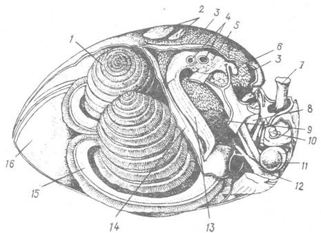
Fig. 146. Internal anatomy of a brachiopod (the dorsal valve and mantle have been removed):
1 — right arm; 2 — dorsal end of the adductor muscles; 3, 4 — hepatic gland lobes and sectioned ducts, respectively; 5 — Stomach; 6 — Heart; 7 — pedicle; 8 — celomoduct; 9 — Blood vessel; 10 — pedicle Muscle; 11 — terminal thickening of the hindgut; 12 — ventral end of the adductor muscle; 13 — Esophagus; 14 — anterior wall of the soft body; 15 — left arm; 16 — ventral shell valve
The permanent shape of the arms is maintained, firstly, by their composition of a robust Cartilage-like tissue; secondly, by the turgor pressure of the fluid within the coelomic canal running through the interior of the arm; and thirdly, by the presence in many brachiopods of specialized skeletal outgrowths of the dorsal shell valve, which are sometimes highly complex in Structure. In some species, muscular and skeletal bridges connect the two arms.
The body of brachiopods is covered by a single-layered epithelium that continues onto the dorsal and ventral sides as a double-layered fold—the mantle—which lines the free portion of the shell not occupied by the soft body, known as the mantle cavity. Along the anterior margin of the mantle, it thickens into a ridge bearing chitinoid marginal setae that protect the filtering apparatus from large debris. Beneath the epithelium lies a thin layer of Connective Tissue, under which rests the ciliated peritoneal epithelium enclosing the voluminous body cavity. Portions of the coelom extend between the two folds of the mantle, forming a rather complex network there, and also penetrate the arms. These include a main coelomic sinus separated from the general coelomic cavity by a partition (the turgor of its coelomic fluid Supports the arms) and an additional, narrower sinus that communicates freely with the former.
In inarticulate brachiopods, the shell valves are joined exclusively by muscles. One unpaired and five paired muscles serve to open and close the valves, slide them anteroposteriorly relative to one another, and move each valve laterally. The pedicle, which emerges between the shell valves, possesses its own musculature. In articulate brachiopods, the valves are opened and closed solely by the action of two groups of paired muscles. The pedicle, which emerges through an opening in the ventral valve, lacks its own muscles. Rotation of the shell around its axis (up to 90°), bending toward the substrate, and returning to a vertical position are accomplished by Muscles Attached to the shell walls and the pedicle.
The Digestive System begins with a small, slit-like mouth located at the Base of the circumoral depression, which is an extension of the ciliated groove of the arms. The depression is covered dorsally by a fold of the integument. The mouth leads into the esophagus, which opens into The Stomach; this is followed by a relatively narrow hindgut that ends blindly in articulate brachiopods, but opens to the outside via an anus in inarticulate ones. Numerous digestive glands (sometimes referred to as hepatic glands) flank the intestine, their ducts converging and emptying into the stomach via four main ducts. Food particles entering these glands from the stomach undergo Digestion AND ABSORPTION. The intestine is looped and suspended within the body cavity by dorsal, ventral, and transverse mesenteries. Brachiopods feed on small planktonic Algae and invertebrates.
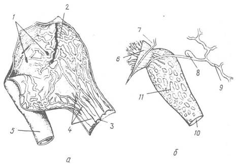
Fig. 147. Stomach opened longitudinally (a) and excretory system (b) of the brachiopod Hemithyris psittacea:
1 — openings of the hepatic gland ducts; 2, 3 — wall of the stomach and hindgut, respectively; 4 — folds on the inner surface of the intestine; 5 — esophagus; 6 — ciliated funnel of the celomoduct; 7 — mesentery; 8 — thickening of the blood vessel (accessory heart); 9 — mantle blood vessel; 10 — excretory pore; 11 — excretory region of the celomoduct
The excretory system is represented by one or two pairs of tubular celomoducts. One end of the celomoduct opens into the coelom via a funnel-shaped expansion, while the narrow opposite end opens externally through a small pore. The excretory function is performed by the tubule of the celomoduct, the inner walls of which have a spongy structure (Fig. 147).
The Circulatory system features a central organ—The Heart—which appears as an elongated muscular sac closely applied to the stomach. A single vessel emerges from the heart, dividing first into two and subsequently into numerous vessels that form ramifications in the mantle and gonad sinuses, supply the celomoducts, and extend into the arms and tentacles. The blood is colorless and lacks cellular elements. Circulation is driven by contractions of The Heart and accessory pulsatile expansions on the vessels, such as those near the celomoducts.

Fig. 148. Development of brachiopods (a–d — Testicardines; e — Ecardines):
a — coelom formation in the embryo; b — free-swimming larva; c — attached larva; d — young brachiopod; e — ecardine larva; 1 — gut rudiment; 2 — coelom; 3 — ocelli; 4 — apical lobe; 5 — muscles; 6 — pedicle rudiment; 7 — setae; 8 — mantle fold; 9 — pedicle; 10 — tentacles; 11 — shell; 12 — preoral lobe
Respiratory Functions are performed primarily by the arms, which provide a large surface area for gas exchange thanks to their numerous thin-walled tentacles.
The Central Nervous system consists of a small paired ganglion located above the esophagus, which is connected by thin connectives to a slightly larger ganglion situated beneath it. The upper ganglion innervates the lophophore arms, while the lower one supplies the rest of the body Organs.
Specialized Sense Organs are absent. In some species, the anterior margin of the mantle bears pigmented light-sensitive spots. Free-swimming brachiopod larvae possess ocelli and statocysts.
All brachiopods are gonochoric, with the exception of three Mediterranean hermaphroditic species of the genus Argyrotheca.
The Gonads (typically two pairs) are located within the coelomic cavities of the dorsal and ventral mantle folds. They are botryoidal in shape; Germ Cells originate beneath the epidermal epithelium and, upon maturation, are released into the coelom, from which they are expelled to the exterior via coelomoducts. Egg Fertilization takes place either within the female mantle cavity (subclass Testicardines) or externally in the water (subclass Ecardines). In the former case, embryonic development proceeds within the mantle cavity either among the folds of the arm epithelium or in a specialized brood pouch formed at the base of the arms, from which a fully formed larva emerges.
Cleavage is total, nearly equal, and approaches radial cleavage, although variations occur. The mesoderm in brachiopods originates differently from that in the preceding phyla—as a pair of pouch-like evaginations of the archenteron (Fig. 148, a) that subsequently pinch off to form two coelomic sacs; this resembles enterocoelous mesoderm formation in deuterostomes (see below).
In articulate brachiopods (subclass Testicardines), a planktonic larva hatches from the egg (Fig. 148, b). Its body comprises three distinct regions: the apical (cephalic), trunk, and pedicle regions. The apical region appears as a ciliated, umbrella-like structure bearing an apical plate and four ocelli; the trunk region features two hanging Skin folds (the dorsal and ventral mantles); and the pedicle region is shaped like a short papilla. The larva is non-feeding; it lacks a mouth and an anus, and the gut is blind-ending. After 10–30 days, the larva settles on the bottom, attaching to the substrate by its pedicle. Both mantle folds then turn upward to enclose the larval trunk and HEAD, with the inner mantle surface becoming the exterior. The sensory apical region regresses, leaving a small fold—the epistome—near the base of which the mouth develops. The mantle folds secrete the shell, and the pedicle region grows into the functional stalk. Rudiments of the arms form near the mouth, initially as two bumps that grow, coil spirally, and develop tentacles (Fig. 148, c, d).
In inarticulate brachiopods (subclass Ecardines), the egg hatches into a larva that already possesses a tentacled lophophore and a bivalved shell (Fig. 148, e); it swims by means of the lophophore, which protrudes from the shell. The larva leads a planktonic, feeding existence for about a month, after which it attaches to the substrate and transforms into an adult brachiopod without undergoing major organ reorganization.
Brachiopods predominantly inhabit the marine shelf zone, with only a small number of species living at considerable depths. Currently, about 30 species are known to occur at depths exceeding 2,000 m, and only three have been found down to 6,000 m. The vast majority of brachiopods securely attach themselves by the ventral valve or pedicle to hard substrates—such as stones, rocks, shells, and other skeletal structures of various invertebrates—while only representatives of the inarticulate family Lingulidae lead a burrowing lifestyle. They inhabit the intertidal and shallow sublittoral zones, excavating vertical mucus-lined burrows in sandy and muddy sediments (Fig. 149). Their long, muscular pedicle anchors in the lower part of the burrow, and upon the slightest disturbance, it contracts rapidly, withdrawing the animal into the burrow. The burrow is constructed and repaired after cave-ins by the digging Movements of the valves and the pedicle.

Fig. 149. Diagram of a representative of the family Lingulidae within its burrow:
a, b — lateral and ventral views, respectively; 1, 2 — animal extended from the burrow and in the retracted state, respectively
Last update: 13/08/2026
Editorial and Educational Adaptation: This material has been compiled based on the primary/original source text. The project team performed an editorial review, corrected technical inaccuracies, structured sections, and adapted the content for an educational format.
What was processed:
- elimination of formatting defects (OCR errors, structural breaks, corrupted characters);
- editorial organization of content;
- standardization of terminology in accordance with academic sources;
- verification of factual statements against the original source text.
All mentions of the author, publication year, and origin of the primary text have been preserved in accordance with the source.