INVERTEBRATE ZOOLOGY IN THREE BOOKS - BOOK 3 - H.Y. Shcherbak - 1997
PHYLUM PHORONIDA
CLASS PHORONIDEA
Phoronids are small to medium-sized animals (the smallest, Phoronis ovalis, is 0.6 cm long, while the largest, Phoronopsis californica, reaches up to 37 cm) that live in tubes made of organic matter, often encrusted with sand grains; these tubes are always longer than the animal's body. Their worm-like body (Fig. 129) bears a horseshoe-shaped projection at the anterior end—the lophophore—with two rows of tentacles, which sometimes spirals as it grows; the posterior end is bulbously swollen (this part is called the ampulla). The number of tentacles depends primarily on the size of the animal, with the largest individuals possessing many hundreds of them.
Phoronids never leave their tubes, within which they move freely, only extending the anterior end of their body with the tentacular apparatus. The body of phoronids is transparent, red, orange, or green, and less frequently colorless.
The body wall is formed by a musculo-cutaneous sac consisting of an epidermal layer covered by a thin cuticle, a basement membrane, a thin outer layer of circular Muscles, and a highly developed inner layer of longitudinal muscles. Internally, the musculo-cutaneous sac is lined by a layer of peritoneal epithelium. The anterior part of the body is the most muscular and contractile.
All muscles in phoronids are smooth.
Scattered among the epidermal Cells are sensory cells associated with the nerve plexus, and glandular cells, some of which secrete the substance from which the tube is constructed.
The general body cavity (coelom) is divided by a Diaphragm into two compartments: that of the tentacular apparatus and that of the trunk. Branches extend from the horseshoe-shaped coelomic channel of the lophophore into all tentacles, reaching the tip of each.
Class="center">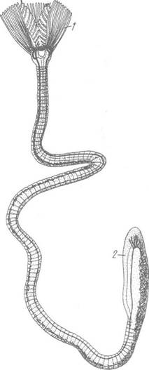
Fig. 129. Phoronis hippocrepia, removed from the tube:
1 — tentacles; 2 — ampulla
The slit-like Mouth is located inside the tentacular crown at the Base of the lophophore and is covered from above by a leathery fold—the epistome. The mouth leads into a ciliated buccal cavity and further into the Esophagus, which transitions into the endodermal Stomach; the latter bends and continues into the narrow section of the midgut, which ascends to the anterior end of the body and opens via the anus outside the tentacular crown (Fig. 130). Phoronids lack an ectodermal hindgut.
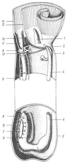
Fig. 130. Diagram of the Internal Structure of phoronids:
1 — lophophore projection; 2 — anus; 3 — coelomoduct; 4 — intestine; 5 — ampulla; 6 — Ovary; 7 — Testis; 8 — Blood vessel; 9 — stomach; 10 — diaphragm; 11 — mouth; 12 — epistome; 13, 14 — inner and outer rows of tentacles
A band of ciliated cells runs along the dorsal wall of The Stomach. Food (small planktonic organisms) filtered from the Water by the tentacular apparatus is glued here into a cord that rotates continuously due to the movement of the cilia. Digestion begins in the stomach cavity and is completed intracellularly within its walls. The intestine is externally covered by peritoneal epithelium and suspended from the body walls by mesenteries.
The excretory system is represented by two V-shaped coelomoducts, each of which opens into the coelom by one or two ciliated funnels, and to the exterior by a single pore. The pores are located on either side of the anus.
The Circulatory system (Fig. 131) is well-developed and almost closed. It consists of vessels and capillaries, with a blood sinus present only around the stomach. The dorsal vessel carries blood to the anterior end of the body, where it transitions into the horseshoe-shaped (venous) vessel of the lophophore; afferent vessels branch from the latter into each of the tentacles. Here, the blood is oxygenated and, via efferent vessels, enters another (arterial), also horseshoe-shaped vessel, from which a second longitudinal, lateral vessel arises, closely adhering to the descending loop of the intestine and carrying arterial blood to the trunk. Numerous capillary branches extend from the lateral vessel, some of which open into the blood sinus in the stomach wall. At the posterior end of the body, both longitudinal vessels merge into each other.

Fig. 131. Diagram of The structure of the CIRCULATORY SYSTEM OF phoronids:
1 — tentacular vessel; 2 — lophophoral vessel; 3, 4 — longitudinal ascending and descending vessels; 5 — branches of vessels to the stomach wall
Phoronids lack a Heart; Blood Circulation is achieved by rhythmic contractions of the walls of the longitudinal vessels, as well as the tentacular vessels and capillaries, which contract autonomously. The blood fluid contains Hemoglobin and other closely related respiratory pigments.
The Nervous system of phoronids is quite primitive. It is based on a diffuse nerve plexus lying within the epithelium above the basement membrane; it has several concentrations: the preoral nerve field, a nerve ring along the lophophore from which nerves innervating the tentacles arise, and a longitudinal nerve cord that extends to the posterior end of the body but does not enter the ampulla. Sensory Cells of the epidermis, which are particularly numerous in the tentacular apparatus, are associated with the nerve plexus.
All known phoronids, with the exception of a few species, are hermaphroditic. The Gonads (ovary and testis) are located in the swollen posterior part of the body. Gametes enter the coelomic fluid and from there pass to the outside via the coelomoducts. Fertilization is external. Fertilized eggs are either immediately dispersed by currents or retained for some time among the tentacles.
Cleavage in phoronids is complete and nearly equal. In some species, it follows a spiral pattern, in others, a radial one. The mouth develops from the anterior part of the blastopore, and the anus forms at the site of its posterior part. The mesoderm arises through the migration of individual cells from the endoderm into the gastrula cavity.
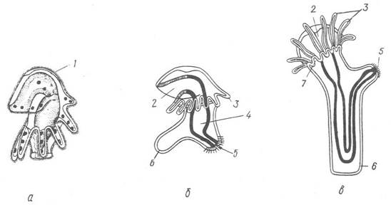
Fig. 132. Development of phoronids:
a — actinotroch; b — evagination of the actinotroch's ventral pocket; c — metamorphosis of the actinotroch; 1 — hood; 2 — mouth;
3 — larval tentacles; 4 — intestine; 5 — anus; 6 — ventral pocket; 7 — primordium of definitive tentacles
The egg hatches into a microscopic planktonic larva (1–5 mm in length) called an actinotroch (Fig. 132). Its upper part, featuring an apical plate, expands significantly to form a hood, beneath which lie the mouth and the larval lophophore with tentacles; the lower part is elongated. A ciliary band runs along the edge of the hood. The actinotroch has a complete gut, a pair of protonephridia, and a primary body cavity containing floating mesodermal cells. On the ventral side of the larva, beneath the lophophore, a sac-like invagination of the ectoderm forms—the ventral pocket, which enlarges as the larva grows. The actinotroch swims in the water Column for 2–3 weeks, feeding on unicellular organisms and detritus particles, before settling to the bottom and finding a suitable substrate for attachment. The subsequent metamorphosis is completed in 15–20 minutes. The actinotroch's ventral pocket evaginates, taking the form of a long cylindrical appendage that projects at a right angle to the rest of the body. This outgrowth grows, the intestinal loop is pulled into it, and it becomes the animal's trunk, while the upper part of the larva, with the mouth and anus, remains at the anterior end, bringing the mouth and anus close together.
The metamorphosis of the actinotroch is somewhat 'catastrophic' in nature: the entire upper part of the larva (the hood with the apical plate and the larval tentacles) is shed; the definitive tentacles of the lophophore develop anew, and the larval protonephridia transform into coelomoducts. The coelom is formed by the settling of mesodermal cells onto the inner wall of the trunk and The surface of the gut; the septum between the two compartments of the coelom forms later.
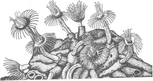
Fig. 133. Aggregation of Phoronis hippocrepia
In phoronids, the autotomy of the anterior end of the body followed by its regeneration is a very common phenomenon. Asexual reproduction in phoronids is also linked to this regenerative capacity. For instance, in Phoronis kowalewskn, a young individual settling on a suitable substrate gives rise to a dense population covering the substrate through multiple transverse fission (architomy). In Phoronis ovalis, the transverse fission of an adult living in a tube produces two individuals. The one formed from the anterior part secretes a transverse septum, separating itself from the other individual, which then creates an opening in the tube next to the septum and builds its own extension tube over it. These divisions result in an entanglement of tubes resembling a true colony.
Most phoronids inhabit the seabed of the coastal zone at depths of up to 50 m in the tropical and temperate zones of all oceans and seas. In the Black Sea, only one species, Phoronis euxinicola, and the actinotroch of another species have been found so far.
Phoronids settle mostly on the shells of dead Mollusks, boring into them. Sometimes they inhabit sandy and muddy sediments, making vertical burrows. As a rule, phoronids live in small aggregations (Fig. 133).

Fig. 133. Aggregation of Phoronis hippocrepia
The geological history of phoronids is unknown. However, based on indirect evidence, some paleontologists consider phoronids to be an ancient group that appeared in the Cambrian–Devonian.
Last update: 13/08/2026
Editorial and Educational Adaptation: This material has been compiled based on the primary/original source text. The project team performed an editorial review, corrected technical inaccuracies, structured sections, and adapted the content for an educational format.
What was processed:
- elimination of formatting defects (OCR errors, structural breaks, corrupted characters);
- editorial organization of content;
- standardization of terminology in accordance with academic sources;
- verification of factual statements against the original source text.
All mentions of the author, publication year, and origin of the primary text have been preserved in accordance with the source.