Review of Medical Physiology - William F. Ganong 2002
Blood Circulation
Regional Blood Circulation
Circulation in Internal Organs
Blood from the intestine, Pancreas, and Spleen flows via the portal vein into the Liver, and from the liver through the hepatic Veins into the INFERIOR VENA CAVA. The Internal Organs and the liver receive 30% of the Cardiac Output via the celiac trunk, as well as the superior and inferior mesenteric Arteries (Fig. 32-15). The liver receives approximately 1000 mL/min from the portal vein and 5000 mL/min from the hepatic artery.
Intestinal Circulation
Blood is supplied to the intestine via several parallel pathways branching from the superior and inferior mesenteric arteries (see Fig. 32-15). Although numerous anastomoses exist between these vessels, occlusion of a major intestinal artery still results in intestinal infarction. Blood flow to the mucosa is greater than to other layers of the intestinal wall and varies with changes in metabolic activity. Consequently, BLOOD FLOW IN the Small Intestine (and, accordingly, in the portal vein) doubles following a meal, remaining elevated for about 3 hours. Intestinal blood flow exhibits well-developed autoregulation.
Class="center">
Fig. 32-15. Splanchnic circulation. Note that most visceral organs receive blood from parallel circuits, whereas the liver receives blood from both the hepatic artery and the portal vein. Mean blood flow is shown in mL/min; C - celiac trunk; SM - superior mesenteric artery; IM - inferior mesenteric artery.
Hepatic Circulation
Large gaps exist between the endothelial Cells of the hepatic sinusoids, resulting in high permeability. The pathway by which the intrahepatic Branches of the hepatic artery and portal vein empty into the sinusoids and subsequently into the central veins is shown in Figures 26-20 and 32-16. The functional unit of the liver is the acinus. Each acinus is centered on a vascular stalk containing terminal branches of the portal vein, hepatic arteries, and Bile ducts. Blood flows from the center of this functional unit toward the terminal hepatic venules at the periphery (see Fig. 32-16). Therefore, the central Regions of the acini, sometimes referred to as zone 1, are well oxygenated, the intermediate zone (zone 2) is moderately oxygenated, and the peripheral zone (zone 3) is the least oxygenated and most vulnerable to anoxic injury. The hepatic veins drain into the inferior vena cava. The acini form cluster-like structures, each on its own stalk. The human liver contains about 100,000 acini. In humans, portal venous pressure normally averages about 10 mmHg, while hepatic venous pressure is about 5 mmHg. The mean pressure in the branches of the hepatic arteries supplying the sinusoids is approximately 90 mmHg; however, pressure within the sinusoids is lower than that in the portal vein, indicating a significant pressure drop across the hepatic arterioles. This pressure drop is maintained by an inverse relationship between blood flow in the hepatic arteries and the portal veins. This reciprocal relationship is mediated by the maintenance of a blood flow level at which adenosine is washed out from the areas surrounding the arterioles. According to this hypothesis, adenosine is produced at a constant rate; as portal blood flow decreases, washout slows down, and the local accumulation of adenosine leads to dilation of the terminal arterioles.
The walls of the intrahepatic branches of the portal vein contain smooth Muscle innervated by noradrenergic vasoconstrictor nerve fibers that enter the liver via the thoracic ventral roots (Th3-Thn) and visceral nerves. Vasoconstrictor Innervation of the hepatic artery is derived from the hepatic sympathetic plexus. No vasodilator fibers supplying the liver have been identified. At rest, blood flow in the peripheral regions of the liver is sluggish, and only a portion of the organ undergoes active perfusion. As systemic venous pressure rises, the branches of the portal vein passively distend, and the blood volume of the liver increases. In congestive Heart Failure, hepatic venous congestion can be pronounced. Conversely, diffuse discharge in noradrenergic fibers in response to a drop in systemic blood pressure causes the intrahepatic branches of the portal vein to constrict, increasing portal pressure and accelerating hepatic blood flow, bypassing much of the organ. The bulk of the blood from the liver enters the systemic circulation. Constriction of the hepatic arterioles diverts blood away from the liver, whereas constriction of the mesenteric arteries reduces portal inflow. In severe Shock, hepatic blood flow may be reduced to a level that causes focal tissue necrosis.
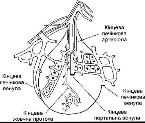
Fig. 32-16. Concept of the acinus as the functional unit of the liver. In each acinus, blood from the portal vein and hepatic arteriole enters the center of the acinus and flows outward toward the hepatic venule (reproduced with permission from Lautt WW, Greenway CV: Conceptual review of the hepatic vascular bed. Hepatology 1987;7:952).
Reservoir Function of the Splanchnic Circulation
In dogs and other mammals, the splenic capsule contains an Abundance of smooth muscle. The spleen traps blood, and rhythmic contractions of its capsule promote the drainage of plasma into Lymphatic vessels. The spleen thus serves as a reservoir for cells-rich blood. Discharges in noradrenergic nerves and adrenaline trigger forceful splenic contractions, discharging blood into the circulation. This splenic function is not quantitatively significant in humans. However, the reservoir function of the entire visceral circulation as a whole is important. For instance, blood accounts for 25–30% of the liver's volume. Contraction of capacitance vessels in visceral organs can mobilize a liter of blood into the Arterial System in less than a minute.
Other reservoirs containing a significant blood volume at rest include the Skin and Lungs. During heavy exercise, vasoconstriction in these organs and the reduction of blood "reserves" in the liver, other splanchnic regions, skin, and lungs can increase the volume of actively perfused circulating blood by 30%.
Last update: 10/08/2026
Editorial and Educational Adaptation: This material has been compiled based on the primary/original source text. The project team performed an editorial review, corrected technical inaccuracies, structured sections, and adapted the content for an educational format.
What was processed:
- elimination of formatting defects (OCR errors, structural breaks, corrupted characters);
- editorial organization of content;
- standardization of terminology in accordance with academic sources;
- verification of factual statements against the original source text.
All mentions of the author, publication year, and origin of the primary text have been preserved in accordance with the source.