Medical Radiology - Lazar A.P. 2008
Radiation therapy
Sources of ionizing radiation
Radiation therapy Methods are divided into two main categories: external beam therapy and brachytherapy.
In external beam radiotherapy, the radiation source is located at a distance from the patient. External beam methods include: teletherapy (external beam gamma therapy), X-ray therapy, high-energy bremsstrahlung therapy, fast electron therapy (beta therapy), and proton therapy.
In contact radiation therapy (also known as brachytherapy), the radiation source is placed directly against or within the pathological focus. This is achieved by placing radioactive Materials On the surface of the irradiated area (surface application), introducing them into a body cavity (intracavitary method), directly into the tumor tissue (interstitial method), or into the patient's body with subsequent accumulation in the affected organ (selective accumulation method).
For the Treatment of certain conditions, simultaneous or sequential combinations of external and contact irradiation are used. This approach is known as combined radiation therapy.
In medical practice, natural and artificial radioactive elements, X-ray units, and various types of particle accelerators serve as sources of ionizing radiation.
They can be classified as follows.
I. Radioactive substances.
1. Sealed sources.
A. Gamma-ray therapy units.
Б. Radioactive sources (tubes, pellets, needles, wires).
2. Unsealed sources (solutions, Suspensions, powders).
II. Charged particle accelerators.
1. Linear accelerators.
2. Cyclic accelerators (betatrons, cyclotrons, synchrophasotrons).
III. X-ray units.
1. Deep therapy (long-distance) units.
2. Contact (short-distance) units.
During radioactive decay, elements emit alpha and beta particles. Alpha and beta decay are typically accompanied by the emission of gamma quanta. Depending on the clinical objective, either beta particles or gamma radiation are utilized for diagnostic or therapeutic purposes in practical medicine.
Depending on their application in medical practice, radioactive substances are divided into two groups: unsealed and sealed radioactive sources.
Unsealed radioactive sources consist of solutions, suspensions, and powders of radioactive elements in various chemical compounds. Artificial radioactive isotopes of iodine (131I), phosphorus (32P), and gold (198Au) have become widely used as unsealed sources in medicine. Because unsealed radiopharmaceuticals carry the risk of spilling, evaporation, scattering, or surface deposition, radiation safety measures for them are more complex, requiring stringent conditions for storage, transport, and handling.
Sealed radioactive sources consist of radioactive elements enclosed in metal capsules shaped as tubes, needles, pellets, discs, wires, etc. The metal casings of sealed sources act as filters that absorb the soft radiation emitted by the element. Each radioactive source is housed in a specific metal cladding; for instance, radium is contained in platinum capsules, while cobalt is kept in stainless steel tubes.
Both natural and artificial elements—such as radium, cobalt, and cesium—are used in sealed sources. Sealed sources also include stationary gamma-therapy units, which utilize artificial radioactive isotopes of cobalt or cesium whose radioactive decay is accompanied by gamma emission.
The most common source of gamma radiation is the radioactive isotope cobalt-60, which has a half-life of 5.24 years and emission energies of 1.17 and 1.33 MeV (averaging 1.25 MeV). To produce this source, a blank made of the stable isotope 59Co is placed in the high-flux region of a nuclear Reactor, where thermal neutron capture leads to the accumulation of radioactive 60Co. These blanks are then assembled to achieve the required total activity and size, placed into stainless steel capsules, and welded shut. Radioactive cobalt can be used in the form of pellets 0.7 mm in diameter and 3 mm in length, each with an activity of 0.3–0.5 mg-eq of radium. To absorb beta radiation, the pellets are coated with a 0.05 mm thick layer of inactive gold. Segments of 60Co are also loaded into nylon tubes with an outer diameter of 1.3 mm and an inner diameter of 0.8 mm, which are used in interstitial radiotherapy. High-activity 60Co sources are utilized for external beam gamma therapy.
Other radioactive sources present both Advantages and disadvantages. For example, 137Cs has a long half-life (30 years) but a low specific activity. Conversely, 192Ir, which is widely used for brachytherapy, has a high activity (3–5 mg-eq of radium)—allowing sources to be manufactured as thin wires (0.6 mm in diameter) and needles—but a short half-life (74 days), necessitating frequent source replacement. The decay of radioactive gold 198Au (T = 2.69 days) is accompanied primarily by beta radiation with a maximum energy of 0.96 MeV and gamma radiation with an energy of 0.41 MeV. Radioactive gold is used as a colloidal solution or in the form of pellets coated with a 0.1 mm thick platinum layer to absorb beta radiation.
A comparative analysis of isodose charts from various radiotherapy machines producing radiation of different natures and energies (Fig. 314) allows for several Conclusions that are crucial for Radiation therapy planning.
X-rays generated at an anode voltage of 30–200 kV cause the maximum absorbed dose to occur at The surface of The Human Body, resulting in the highest exposure to the Skin. At greater tissue depths, the dose decreases significantly. At an anode voltage of 40 kV, the dose at a depth of 3 cm is only 10% of the surface dose. At an anode voltage of 200 kV, the radiation penetrates deeper, yet even here, only 20% of the surface dose remains at a depth of 10 cm. Therefore, X-ray units are used exclusively for irradiating superficial tumors.
Class="center">
Fig. 314. Dose distribution in Tissues for various Types of ionizing radiation:
X - X-ray;
Y - gamma;
Г - bremsstrahlung;
Е - electron;
П - proton;
Н - neutron.
By adjusting the voltage and filters of the X-ray machine, radiation of varying penetrating power can be generated. Long-distance X-ray therapy is performed at a source-to-skin distance of more than 30 cm (Fig. 315). The current remains constant at approximately 10 mA. Superficial X-ray therapy is used when the pathological focus is located at a depth of up to 1 cm from the skin surface; it employs a tube voltage of 100–120 kV, a 3 mm thick aluminum filter, and an SSD of 30 cm. Semi-deep X-ray therapy is used when the lesion is located at a depth of up to 3 cm; it employs a voltage of 140–160 kV, a 0.5 mm thick copper filter combined with a 3 mm thick aluminum filter, and an SSD of 40–50 cm. Deep X-ray therapy is used for lesions located at a depth of up to 5 cm; it employs a voltage of 180–230 kV, a 1.0 mm thick copper filter combined with a 5.0 mm thick aluminum filter, and an SSD of 40–50 cm. To limit the irradiation field to the required size and shape, lead-walled specialized cones (tubuses) are installed in the exit window of the X-ray tube (Fig. 316).

Fig. 315. RUM-11 X-ray therapy unit.
For short-distance X-ray therapy, therapy units with a remote anode in the form of a cylindrical rod are used. The remote anode can also be employed for intracavitary therapy. The source-to-surface distance in short-distance X-ray therapy ranges from 1.5 to 30 cm (most commonly 10 cm), and the tube voltage is regulated between 8 and 100 kV. Short-distance X-ray therapy is applied when the pathological focus is located at a depth of up to 0.5–0.7 cm from the skin surface. A variant of short-distance X-ray therapy is ultra-soft X-ray therapy, in which so-called Bucky rays are generated at voltages ranging from 10 to 20 kV. The penetrating power of Bucky rays into human body tissues does not exceed 1.5 mm. These rays are used to treat hyperkeratosis and superficially located inflammatory processes such as dermatitis, blepharitis, and Conjunctivitis.
Gamma-therapy units loaded with 60Со with an activity of 2,000–4,000 Ci emit a nearly uniform beam of high-energy photons (1.25 MeV). The absorption maximum lies at a depth of 0.5 cm, which reduces skin exposure. At a depth of 10 cm, at least 50% of the surface dose remains. Consequently, deep-tissue doses are higher than those achieved with conventional X-ray therapy units.

Fig. 316. Set of filters and cones for X-ray therapy.

Fig. 317. Gamma-therapy unit.
A - external view; Б - Structure OF THE radiation HEAD;
1 - shutter;
2 - Diaphragm;
3 - 60Со source.
External beam gamma therapy units such as AGAT-R, Rokus, and Theratron feature a radiation head equipped with a collimator that regulates the irradiation field size and a light localizer. The radiation head can rotate around the patient, facilitating patient positioning and beam centering (Fig. 317). The units are supplied with a set of removable parts, including a kit of shielding and wedge blocks with a rotating mechanism, lattice filters and grids with varying ratios of blocked and open areas, and additional collimators that allow for a wide range of γ-irradiation techniques in treating various malignant neoplasms (Figs. 318, 319).

Fig. 318. Wedge blocks. 1 - various types of blocks; 2 - external beam gamma therapy for esophageal Cancer:
A - without blocks;
B - with wedge filters.

Fig. 319. Additional filters and blocks for teletherapy (external beam radiotherapy).
A - grid filters;
B - shielding blocks.

Fig. 320. Linear accelerator.
Linear electron accelerators (LEAs) and cyclic accelerators (betatrons and microtrons) producing bremsstrahlung and electron beams are increasingly used to treat patients with malignant tumors (Fig. 320). Compared to gamma-ray units, accelerators offer several advantages, including the choice of radiation type (photon or electron), adjustable radiation energy, and a remarkably small electron beam size (up to 0.5 mm).
In betatrons, the deceleration of the electron stream generates bremsstrahlung with energies ranging from 4 to 40 MeV. At a photon energy of 25 MeV, the maximum absorbed dose occurs at a depth of 4–6 cm beneath the patient's skin. Tissues located at this depth receive no more than half of the maximum dose. However, bremsstrahlung has a notable drawback—a relatively slow dose fall-off beyond the maximum. This means that healthy tissue situated behind the tumor receives significant radiation.
When high-energy electron beams are produced in a betatron, the peak absorbed dose is located at a depth of 1–3 cm, after which the dose drops sharply, causing virtually no radiation to surrounding tissue at a depth of 10 cm. This makes it highly effective for treating superficial neoplasms. However, for deep-seated tumors, heavy charged particle beams (protons and alpha particles) are the preferred choice.
Proton radiation is a type of ionizing radiation consisting of heavy charged particles known as protons. Medical proton beams are generated using large particle accelerators. The energy of proton beams used in radiation therapy typically ranges from 50 to 1000 MeV. The main advantage of proton radiation over gamma, electron, and bremsstrahlung radiation is that high-energy protons undergo very little scattering as they pass through tissues, allowing for the selective destruction of tumors located in close proximity to vital Organs. Typically, protons with energies of 70–200 MeV are used for irradiation. High-energy protons travel in an almost straight line until they "stop" in the tissue, with linear energy transfer (LET) increasing and reaching its maximum at the end of their range (the Bragg peak).
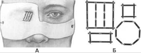
Fig. 321. Surface (applicator) brachytherapy.
A - facial application using linear radioactive sources;
B - layout diagrams of linear radioactive sources within a mold.
A medical proton facility consists of a proton beam transport system, a treatment room housing some of the magnetic focusing elements, a beam monitoring system, and equipment for proton stereotactic radiosurgery. The optimal application of this method is radiosurgery, which involves bloodless Procedures on various areas of the Brain. The ability to shape a narrow beam delivering a high radiation dose (100–200 Gy) makes it possible to selectively destroy brain volumes of 1 cm or more, such as pituitary tumors.
Contact methods of radiation therapy (brachytherapy) include surface (applicator), intracavitary, interstitial, and internal (or targeted accumulation) methods.
Surface brachytherapy employs applicators (derived from the Latin "applicare" - to attach). Plastics, polyethylene films, and ion-exchange resins containing radionuclides are used as applicators (Fig. 321). These are custom-shaped and applied directly to the pathological site. Both beta and gamma applicators are used. Beta applicators, containing the radionuclides Sr90 and Y90, are widely used in ophthalmology. Irradiation is delivered through the working surface of the applicators placed against the pathological focus on an outpatient basis, or surgically secured for a specified period of time.
When radiation sources are placed inside a natural body cavity (such as the Esophagus, Urinary Bladder, Vagina, Uterus, or rectum), the Procedure is called intracavitary irradiation (Fig. 322). High-activity 60Co, 137Cs, and 192Ir gamma-ray sources are widely used to treat cancers of the uterine cervix and corpus. Special computer-controlled afterloading systems are used for intracavitary and surface irradiation, such as the "AGAT-VU" (loaded with 60Co), "Selectron" (loaded with 137Cs), and "Gammamed" (loaded with 192Ir). These devices remotely transport radiation sources through flexible tubes into endostats previously positioned within the cavity (Fig. 323). This technique is known as remote afterloading.

Fig. 322. Intracavitary irradiation.
A - linear radiation sources within the uterine cavity;
B - dose distribution using a linear source.
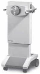
Fig. 323. GammaMed apparatus.
Interstitial irradiation using sealed sources is performed by inserting wire, needle, or tube-like guides into the tumor, into which radiation sources—most commonly 60Co—are subsequently placed (Fig. 324). Typically, during the procedure, a visual placement of the guides is performed, after which radioactive sources are introduced using specialized equipment («MicroSelectron»). A 125I radioactive source with an activity of 0.8224 mCi can remain within the tumor for up to 6 months.
The radiosurgical method, as a variant of the interstitial approach, involves the surgical removal of the tumor followed by the administration of radioactive agents into the Tissues of the tumor bed. Radioactive agents, needles, or nylon tubes are arranged in rows within the soft tissues of the wound. To prevent displacement, each of them is secured with a single catgut suture. Nylon threads do not require additional fixation since the tissues are sutured directly with them. After administering the radioactive agents, the wound is closed, and the threads from each agent or the ends of the nylon tubes are brought out through the gaps between the sutures. Upon completion of the exposure, after 5-7 days, the devices are removed. The total dose can range from 45 to 60 Gy, depending on the radicality of the surgery. Intraoperative Electron Beam Radiotherapy (IOERT) can also be applied during the procedure (Fig. 325).
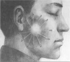
Fig. 324. Interstitial irradiation of a parotid gland tumor.

Fig. 325. Intraoperative beta-irradiation.
1, 3 - delivery of irradiation; 2 - irradiation scheme on a computed tomography scan.
Irradiation with unsealed liquid radionuclides (internal method) is carried out by directly introducing a radioactive substance into the body as a true or colloidal solution orally, into a cavity, tumor, or vessel (Fig. 326). The Use of aqueous solutions of 32P and 131I salts is based on their ability to selectively accumulate primarily in specific tissues (phosphorus in the Bone Marrow, iodine in The Thyroid Gland) regardless of the route of administration; therefore, these radionuclides are used to treat Blood disorders and thyroid cancer with metastases. Colloidal solutions of 198Au are more frequently used for intracavitary, intratumoral, and intralymphatic administration. To administer radioactive gold into the area of the surgical scar, long hollow needles are inserted beforehand, after which a syringe cannula containing the colloidal gold solution is connected to the needle. The solution is injected while simultaneously and gradually withdrawing the needle.
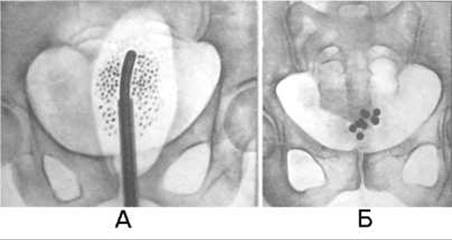
Fig. 326. Intracavitary irradiation on radiographs.
A - balloon with a macrosuspension of the radioactive agent in the rectum;
B - radioactive cobalt spheres in the urinary bladder.

Fig. 327. Stereotactic device.
Last update: 08/08/2026
Editorial and Educational Adaptation: This material has been compiled based on the primary/original source text. The project team performed an editorial review, corrected technical inaccuracies, structured sections, and adapted the content for an educational format.
What was processed:
- elimination of formatting defects (OCR errors, structural breaks, corrupted characters);
- editorial organization of content;
- standardization of terminology in accordance with academic sources;
- verification of factual statements against the original source text.
All mentions of the author, publication year, and origin of the primary text have been preserved in accordance with the source.