Human Anatomy - Kotsan I. Y. 2009
Splanchnology
Digestive System
Stomach
The Stomach (ventriculus, gaster) is an expanded section of the digestive tract located between the Esophagus and the duodenum. It serves as a food reservoir, mixes food and moves it into the Small Intestine, and processes it chemically through the secretion of gastric juice, which contains Pepsin, lipase, Hydrochloric acid, and mucus. The stomach also performs bacteriological and absorptive Functions (absorbing Water, sugars, salts, and alcohol). Furthermore, the gastric mucosa produces anti-anemic substances (Castle's factors)—complex compounds that influence hematopoiesis.
The stomach is situated in the upper Abdominal cavity beneath the Diaphragm and Liver. Three-quarters of the organ lie in the left hypochondriac region, while one-quarter is located in the epigastric region. The cardiac opening of the stomach is at the level of the eleventh thoracic vertebra, and the pyloric opening is at the level of the first lumbar vertebra. The anterior surface of the stomach contacts the diaphragm, the left lobe of the liver, and the anterior abdominal wall. Posterior to the stomach lie the Pancreas, Spleen, transverse colon with its mesentery, left Adrenal gland, upper segment of the left Kidney, and the Abdominal Aorta.
The shape, position, and dimensions of the stomach are highly variable and depend on numerous factors. In a living person, three main stomach shapes are distinguished based on body build types. In individuals with a brachymorphic body type, the stomach is horn-shaped; in mesomorphic individuals, it resembles a fishhook; and in dolichomorphic individuals, it has the shape of an elongated stocking. These shapes, as well as many intermediate variants, are observed when the person is in an upright position. When lying on the back or side, the shape of the stomach changes primarily due to alterations in its spatial relationships with neighboring Organs. The shape of the stomach also depends on sex and age: women more frequently have an elongated hook-shaped stomach, whereas the elderly and children tend to have a horn-shaped stomach.
The size of the stomach, like its shape, varies considerably both individually and depending on the degree of its filling. An empty stomach is small in size and resembles the intestine with a slight expansion in the region of its fundus and body. With moderate filling, the stomach assumes a pear shape, with an average capacity in adults of about 3 L. Upon significant distension, its capacity increases to 4–5 L, and the stomach itself descends to the level of the umbilicus or even lower (typically, the lower border of the stomach is projected 3 cm above the umbilicus). The length of an empty stomach is 18–20 cm, and the distance between the greater and lesser curvatures is 7–8 cm. At moderate filling, the stomach length is 24–26 cm, and the maximum distance between the greater and lesser curvatures does not exceed 10–12 cm.
The stomach presents an anterior wall (paries anterior), facing forward, slightly upward, and to the right, and a posterior wall (paries posterior), facing backward, downward, and to the left. At the margins of the stomach, where one wall transitions into the other, the lesser curvature of the stomach (curvatura ventriculi minor) is formed, directed to the right and upward, along with the greater curvature of the stomach (curvatura ventriculi major), directed to the left and downward (Fig. 147). The lesser curvature of the stomach is concave and short. At the upper part of the lesser curvature lies the cardiac orifice (ostium cardiacum), which is the site where the esophagus enters the stomach. In the lower section of the lesser curvature, there is an indentation called the angular notch (incisura angularis). The greater curvature is convex and long, terminating above at the cardiac notch (incisura cardiaca), near the junction of the esophagus and stomach.
Class="center">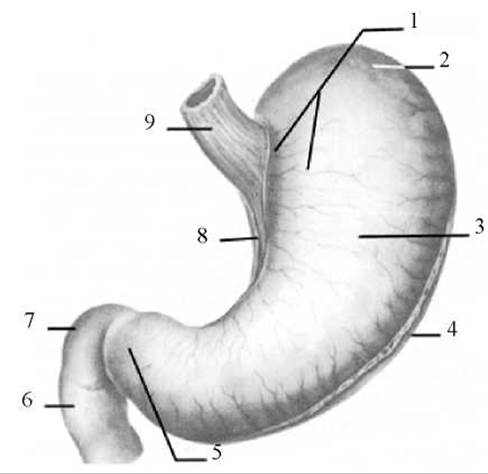
Fig. 147. Stomach (anterior view)
1 — cardiac (inlet) part; 2 — fundus (fornix) of the stomach; 3 — body of the stomach; 4 — greater curvature of the stomach; 5 — pyloric part; 6 — descending part of the duodenum; 7 — superior part of the duodenum; 8 — lesser curvature of the stomach; 9 — esophagus
The stomach is subdivided into the cardiac part (pars cardiaca), where the esophagus opens; the fundus (fornix) of the stomach (fundus (fornix) ventriculi), which is the upwardly convex region adjacent to the left half of the diaphragm; the body of the stomach (corpus ventriculi), which is the largest portion lying between the cardiac and pyloric parts; and the pyloric part (pars pylorica), which is the lower narrowed division of the stomach. Within the pyloric part, a wider region called the pyloric antrum (antrum pyloricum) and a narrower pyloric canal (canalis pyloricus) leading to the duodenum are distinguished. The boundary between the stomach and the duodenum is marked by a circular groove known as the pylorus (pylorus), which corresponds to the exit opening of the stomach termed the pyloric orifice (ostium pyloricum).
The wall of the stomach consists of an outer serous coat, a subserous layer, a muscular coat, a submucous layer, and a mucous membrane (mucosa).
The serous coat (tunica serosa) of the stomach is the visceral Peritoneum, covering the organ on all sides, meaning the stomach is positioned intraperitoneally. Only narrow strips along the greater and lesser curvatures remain uncovered. In these areas, the two peritoneal layers pass from the anterior and posterior walls of the organ onto adjacent organs, forming the peritoneal ligaments of the stomach: the hepatogastric ligament (lig. hepatogastricum), the gastrocolic ligament (lig. gastrocolicum), and the gastrosplenic ligament (lig. gastrosplenicum). Blood Vessels and nerves run to the stomach within the thickness of these ligaments (between the peritoneal layers). These ligaments, along with the relatively fixed portions of the stomach around its inlet and outlet orifices, serve as the fixation factors of the stomach. A delicate subserous layer (tela subserosa) separates the serous coat from the muscular coat.
The muscular coat (tunica muscularis) is the thickest of all the stomach layers. It is composed of three layers of smooth Muscle fibers: an outer longitudinal layer, a middle circular layer, and an inner oblique layer.
The longitudinal layer (stratum longitudinale) is a continuation of the corresponding muscular layer of the esophagus. It is best developed in the region of the greater and lesser curvatures of the stomach.
The circular layer (stratum circulare) is more strongly developed than the longitudinal layer and encircles the stomach along its entire length. In the pyloric region of the organ, this layer thickens to form the pyloric sphincter muscle (m. sphincter pylori) near the exit opening of the stomach.
The oblique fibers (fibrae obliquae) are present only in the region of the body and Cytology/practical/108.html">Fundus of the stomach. They arch over the cardiac part of the stomach to the left of the inlet orifice and pass obliquely downward and to the right across the anterior and posterior walls of the organ toward the greater curvature.
Such a diverse orientation of fibers ensures a variety of movements within the stomach that promote thorough mixing of food with gastric juice.
The muscular coat is separated from the mucosa by a well-defined submucous layer, which allows the overlying mucosa to gather into gastric folds (plicae gastricae).
The mucous membrane (tunica mucosa) of the stomach is lined with a single-layered columnar Glandular Epithelium that secretes mucus. The thickness of this membrane is 2–3 mm. Due to the presence of the muscularis mucosae and a well-developed submucous layer, the mucous membrane forms numerous gastric folds of varying length and direction (Fig. 148). Specifically, long longitudinal folds are located near the lesser curvature, while transverse, oblique, and longitudinal folds are found in the region of the fundus and body, with the long folds interconnected by short ones. The direction and number of folds are more or less constant. When the stomach is distended, the mucosal folds flatten out.

Fig. 148. Gastric mucosa (internal surface of the posterior wall)
1 — muscular coat of the esophagus; 2 — cardiac notch of the stomach; 3 — fundus of the stomach; 4 — folds of the gastric mucosa; 5 — mucous membrane; 6 — submucous layer; 7 — muscular coat of the stomach; 8 — pyloric valve; 9 — pyloric sphincter muscle; 10 — pyloric (outlet) part; 11 — angular notch; 12 — folds of the gastric mucosa; 13 — cardiac (inlet) part; 14 — cardiac orifice; 15 — folds of the esophageal mucosa.
At the transition of the stomach into the duodenum, the pyloric mucosa forms a circular fold known as the pyloric valve. The pyloric valve (valvula pylorica) bounds the pyloric orifice and, upon contraction of the pyloric sphincter muscle, completely isolates the gastric cavity from the duodenum.
The entire surface of the gastric mucosa (on and between the folds) features small elevations (1–6 mm in diameter) called gastric areas (areae gastricae). The surface of these areas contains depressions known as gastric pits (foveolae gastricae), which represent the openings of numerous (approximately 35 million) tubular gastric glands. Three groups of glands are distinguished: cardiac glands (glandulae cardiacae); proper gastric glands (glandulae gastricae propriae), located in the region of the fundus and body of the stomach and consisting of chief and parietal Cells; and pyloric glands (glandulae pyloricae), which consist of chief cells. Chief cells produce pepsinogen, an enzyme of the gastric juice, while parietal cells secrete hydrochloric acid and the intrinsic anti-anemic factor. In the presence of hydrochloric acid, pepsinogen digests food Proteins. Additionally, the gastric mucosa contains cells resembling chief cells but much smaller—so-called mucous neck cells. These cells produce a mucoid secretion (mucus) and are found in the mucosa across all PARTS OF THE stomach. Lymphoid nodules are scattered throughout the mucosa in places, with their primary aggregations located in the pyloric region.
Blood supply to the stomach is provided by the left gastric artery (a branch of the celiac trunk) and the right gastric artery (a branch of the hepatic artery proper), which approach the lesser curvature of the stomach, as well as by the left gastro-omental artery and short gastric Arteries (Branches of the splenic artery) and the right gastro-omental artery (a branch of the gastroduodenal artery), which supply the greater curvature. The gastric and gastro-omental arteries anastomose with one another along the lesser and greater curvatures, forming a ring around the stomach from which numerous branches extend into the gastric walls. Venous blood drains via similarly named Veins into the portal vein and its tributaries.
The lymphatic bed of the stomach is well developed and consists of intramural capillaries and vessels, as well as extra-organ draining Lymphatic vessels. The efferent lymphatic vessels run to the gastric (right and left), gastro-omental (right and left), hepatic, pancreaticosplenic, celiac, diaphragmatic, and mediastinal Lymph Nodes.
The Innervation of the stomach is provided by branches of the Vagus nerve and the Sympathetic trunk. The former increases glandular secretion, enhances peristalsis, and relaxes the pyloric sphincter, while the latter acts as its antagonist.
Last update: 08/08/2026
Editorial and Educational Adaptation: This material has been compiled based on the primary/original source text. The project team performed an editorial review, corrected technical inaccuracies, structured sections, and adapted the content for an educational format.
What was processed:
- elimination of formatting defects (OCR errors, structural breaks, corrupted characters);
- editorial organization of content;
- standardization of terminology in accordance with academic sources;
- verification of factual statements against the original source text.
All mentions of the author, publication year, and origin of the primary text have been preserved in accordance with the source.