Human Anatomy - Kotsan I. Y. 2009
Splanchnology
Digestive System
Oral Cavity
The Oral Cavity (cavitas oris) is the initial section of the digestive tract (Fig. 140). Equipped with general and taste receptors, it assesses food quality; Teeth grind the food, while the Tongue mixes it with saliva coming from the Salivary Glands, and then directs it into the Pharynx.
Class="center">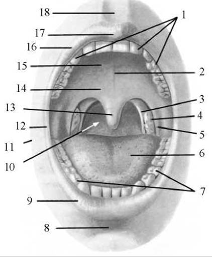
Fig. 140. Oral Cavity and fauces
1 — upper dental arch; 2 — palatine raphe; 3 — palatopharyngeal arch; 4 — palatine tonsil; 5 — palatoglossal arch; 6 — dorsum of the tongue; 7 — lower dental arch; 8 — mentolabial sulcus; 9 — lower lip; 10 — ISTHMUS OF THE fauces; 11 — cheek; 12 — labial commissure; 13 — uvula; 14 — soft palate; 15 — hard palate; 16 — upper lip; 17 — tubercle of the upper lip; 18 — philtrum
The oral cavity is located in the lower part of the face. It is bounded inferiorly by the Suprahyoid Muscles, which form the floor of the Mouth, and superiorly by the Hard and Soft palate. The Cheeks form its lateral boundaries, the Lips form the anterior boundary, and posteriorly, through an opening called the fauces, the oral cavity connects to the pharynx. The teeth and tongue are located within the oral cavity, and the ducts of the Major and minor salivary glands open into it.
The oral cavity is divided into the Vestibule of the mouth and the oral cavity proper (Fig. 141).
The vestibule of the mouth (vestibulum oris) is a narrow cleft bounded anteriorly and laterally by the lips and cheeks, and internally by the teeth and Gums. The vestibule communicates with the external environment via the oral fissure, and with the oral cavity proper through the gaps between the upper and lower teeth as well as the openings located posterior to the last teeth of the Mandible.
The oral fissure (rima oris) is bounded by the upper and lower lips (labium superius et labium inferius). The structural foundation of the lips is formed by the fibers of the orbicularis oris Muscle. The outer surface of the lips is covered with Skin, while the inner surface is lined with mucous membrane. The intermediate zone (the red vermilion border) of the lips is covered with non-keratinized stratified squamous epithelium. The mucous membrane of the lips and cheeks continues onto the alveolar processes of the Maxilla and mandible, forming the gums, which firmly attach to the alveolar margins of the jaws and the necks of the teeth. Along the midline, between the lips and the gums, the mucous membrane thickens to form two folds: the frenulum of the upper lip and the frenulum of the lower lip (frenulum labii superioris and frenulum labii inferioris). The upper lip transitions into the lower lip at the corners of the mouth, forming the labial commissure (commissura labiorum). On the anterior surface of the upper lip, right in the middle, There is a small elevation known as the tubercle (tuberculum). The groove extending along the midline from the tubercle to the nasal septum is called the philtrum (this name originated from the sweat droplets that tend to accumulate more densely in this depression on a perspiring face). Numerous mucous labial glands are located in the submucosal tissue of the lips, with their excretory ducts opening onto the mucosal surface of both lips.

Fig. 141. Oral cavity and pharyngeal cavity (sagittal section)
1 — oral cavity proper; 2 — vestibule of the mouth; 3 — inferior nasal meatus; 4 — nasal vestibule; 5 — frontal sinus; 6 — middle nasal concha; 7 — Inferior nasal concha; 8 — superior nasal concha; 9 — sphenoid sinus; 10 — pharyngeal tonsil (adenoid); 11 — pharyngeal opening of the auditory tube; 12 — torus tubarius; 13 — soft palate (palatine velum); 14 — oral part of the pharynx (oropharynx); 15 — palatine tonsil; 16 — isthmus of the fauces; 17 — ROOT of the tongue (lingual tonsil); 18 — epiglottis; 19 — aryepiglottic fold; 20 — laryngeal part of the pharynx (laryngopharynx); 21 — cricoid Cartilage; 22 — Esophagus; 23 — Trachea; 24 — thyroid cartilage; 25 — Hyoid bone; 26 — geniohyoid muscle; 27 — genioglossus muscle; 28 — mandible
The cheeks (buccae), much like the lips, are covered externally by skin and internally by a mucous membrane. The structural Base of the cheek is formed by the buccinator muscle. Between the skin and the buccinator muscle lies a depression containing a prominent mass of adipose tissue—the buccal fat pad (corpus adiposum buccae). This pad is particularly well-developed in infants during their first years of life, giving their faces a characteristic rounded appearance. These structures are believed to play a special role in newborns by helping to reduce atmospheric pressure within the oral cavity during suckling. A large number of minor salivary glands, as well as the ducts of the parotid glands, open into the oral vestibule. The orifice of the parotid duct is located on the buccal mucosa opposite the upper second molar.
The oral cavity proper (cavitas oris propria) is bounded anteriorly and laterally by the gums and teeth, superiorly by the hard and partially soft palate, and inferiorly by the floor of the mouth, which is formed by the paired mylohyoid and geniohyoid muscles. Posteriorly, the oral cavity communicates with the pharynx through the fauces—an unpaired opening bounded superiorly by the soft palate (its palatine velum and uvula), inferiorly by the root of the tongue, and laterally by the palatine arches.
The palate (palatum) consists of two parts: an anterior (larger) part—the hard palate, and a posterior (smaller) part—the soft palate.
The hard palate (palatum durum) separates the oral cavity from the Nasal cavity. Its bony framework is formed by the palatine processes of the maxillae and the horizontal plates of the palatine bones. The overlying mucous membrane is thick, smooth, and tightly fused with the periosteum. In the anterior region of the palate, there are several faint transverse palatine folds (plicae palatinae transversae), while the median line features the palatine raphe (raphe palati), which marks the line of fusion of the maxillae.
The soft palate (palatum molle) is composed of a fibrous sheet and muscles. It comprises an anterior horizontal part, which is a continuation of the hard palate, and a posterior part that extends obliquely backward and downward, known as the palatine velum (velum palatinum). In the midline, the palatine velum extends into a small, cone-shaped process called the uvula (uvula palatina). On either side, the palatine velum continues into two arches: the anterior palatoglossal arch (arcus palatoglossus) and the posterior palatopharyngeal arch (arcus palatopharyngeus), between which the palatine tonsil (tonsilla palatina) is lodged.
Beneath its mucous membrane, the soft palate contains a Connective Tissue plate known as the palatine aponeurosis, as well as a set of muscles that play a crucial role in the act of swallowing.
The musculus uvulae (m. uvulae) originates from the posterior nasal spine of the Palatine bone and inserts into the substance of the tongue. Function: elevates the uvula and draws it backward.
The levator veli palatini muscle (m. levator veli palatini) originates from the Inferior surface of the petrous part of the Temporal bone and attaches to the palatine aponeurosis. Function: elevates the soft palate and pulls it posteriorly, and widens the cartilaginous part of the auditory tube, thereby improving communication between the tympanic cavity and the nasopharynx.
The tensor veli palatini muscle (m. tensor veli palatini) arises from the Sphenoid bone and inserts into the palatine aponeurosis. Function: tenses the palatine velum and the cartilaginous part of the auditory tube, increasing its lumen.
The palatoglossus muscle (m. palatoglossus) runs within the substance of the palatoglossal arch. It originates from the lateral margin of the root of the tongue and attaches to the palatine aponeurosis. Function: tenses the arches and thereby narrows the fauces.
The palatopharyngeus muscle (m. palatopharyngeus) originates from the posterior wall of the pharynx and the lamina of the thyroid cartilage, and inserts among the muscle bundles of the soft palate and its aponeurosis. Some bundles of this muscle attach to the lower margin of the medial lamina of the auditory tube cartilage, forming the salpingopharyngeus muscle. Function: approximates the palatopharyngeal arches (narrowing the opening of the fauces) and pulls the lower part of the Pharynx and Larynx upward.
Last update: 08/08/2026
Editorial and Educational Adaptation: This material has been compiled based on the primary/original source text. The project team performed an editorial review, corrected technical inaccuracies, structured sections, and adapted the content for an educational format.
What was processed:
- elimination of formatting defects (OCR errors, structural breaks, corrupted characters);
- editorial organization of content;
- standardization of terminology in accordance with academic sources;
- verification of factual statements against the original source text.
All mentions of the author, publication year, and origin of the primary text have been preserved in accordance with the source.