Human Anatomy and Physiology - N. I. Fedyukovich 2003
Cardiovascular system
Lymphatic system
The Lymphatic system is a part of The Cardiovascular system (Fig. 97). It returns Water, Proteins, Lipids, and metabolic products from Tissues to the bloodstream.
Class="center">
Fig. 97. The lymphatic system (diagram):
1,2 — parotid Lymph Nodes; 3 — cervical nodes; 4 — Thoracic duct; 5, 14 — axillary lymph nodes; 6, 13 — cubital lymph nodes; 7, 9 — inguinal lymph nodes; 8 — superficial lymphatic Vessels of the lower leg; 10 — iliac nodes; 11 — mesenteric nodes; 12 — cisterna chyli; 15 — subclavian nodes; 16 — occipital nodes; 17 — submandibular nodes
The lymphatic system performs several Functions: 1) maintaining the volume and composition of interstitial fluid; 2) maintaining humoral communication between the interstitial fluid of all Organs and tissues; 3) absorption and transport of nutrients from the digestive tract into the Venous system; 4) transporting migrating lymphocytes and plasma Cells to the Bone Marrow and sites of injury. The lymphatic system also transports malignant tumor cells (metastases) and microorganisms.
The human lymphatic system consists of lymphatic vessels, lymph nodes, and lymphatic ducts.
The lymphatic system originates from lymphatic capillaries. They are present in all organs and Tissues of the human body, except for the Brain AND SPINAL cord and their Meninges, the Skin, the Placenta, and the splenic parenchyma. The capillary walls are thin, single-layered endothelial tubes with a diameter of 10 to 200 μm, ending blindly. They are highly distensible and can expand 2 to 3 times their normal size.
The fusion of several capillaries forms a lymphatic vessel, which also contains the first valve. Depending on their depth, lymphatic vessels are divided into superficial and deep. Lymph flows through these vessels to the lymph nodes corresponding to a specific organ or body region. Depending on the source of the lymph, lymph nodes are classified as visceral, somatic (parietal), or mixed. The first group drains lymph from Internal Organs (such as tracheobronchial nodes); the second drains The Musculoskeletal System (popliteal, cubital nodes); the third drains the walls of hollow organs; and the fourth drains deep body structures (deep cervical nodes).
Vessels that carry lymph to a node are called afferent lymphatic vessels, while those leaving the hilum of the node are called efferent lymphatic vessels.
Large lymphatic vessels form lymphatic trunks, which merge to form lymphatic ducts that empty into venous angles or the terminal segments of the Veins that form them.
There are six such large lymphatic ducts and trunks in The Human Body. Three of them (the thoracic duct, the left jugular trunk, and the left subclavian trunk) empty into the left venous angle, while the other three (the Right lymphatic duct, the right jugular trunk, and the right subclavian trunk) empty into the right venous angle.
The thoracic duct is formed retroperitoneally in the Abdominal cavity at the level of the XII thoracic and II lumbar vertebrae by the fusion of the right and left lumbar lymphatic trunks. It is 20–40 cm long and drains lymph from the lower limbs, the walls and Organs of the pelvis, the abdominal cavity, and the left half of the chest. From the abdominal cavity, the thoracic duct passes through the aortic hiatus into the thoracic cavity, then enters the neck region and opens into the left venous angle or the terminal segments of its forming veins. The bronchomediastinal trunk, which drains lymph from the left half of the chest, empties into the cervical part of the duct; the left subclavian trunk carries lymph from the left arm; and the left jugular trunk comes from the left side of the HEAD and Neck. Along the course of the thoracic duct, there are 7–9 Valves that prevent the backflow of lymph.
The right lymphatic duct drains lymph from the right side of the head, neck, upper limb, and organs of the right half of the chest. It is formed by the right subclavian, right bronchomediastinal, and right jugular trunks, and empties into the right venous angle.
The Lymphatic vessels and nodes of the lower limb are divided into superficial and deep. Superficial vessels drain lymph from the skin and subcutaneous tissue of the FOOT, lower leg, and thigh. They empty into the superficial inguinal lymph nodes located below the inguinal ligament. Lymph from the anterior abdominal wall, gluteal region, external genitalia, Perineum, and some pelvic organs also drains into these nodes.
The popliteal lymph nodes are located in the popliteal fossa and drain lymph from the skin of the foot and lower leg. The efferent vessels of these nodes empty into the deep inguinal lymph nodes.
Deep lymphatic vessels drain lymph from the foot and lower leg into the popliteal lymph nodes, and from the tissues of the thigh into the deep inguinal nodes, whose efferent vessels drain into the external iliac nodes.
Depending on their Location, the pelvic lymph nodes are divided into parietal and visceral. The first group includes the external, internal, and common iliac nodes, which drain lymph from the pelvic walls. Visceral lymph nodes, located near the pelvic organs, include paravesical, parametrial, paravaginal, and pararectal nodes, draining lymph from their respective organs.
The efferent vessels of the internal and external iliac nodes reach the common iliac lymph nodes, from which lymph flows to the lumbar nodes.
Lymph is collected in the abdominal lymph nodes from the parietal and visceral LYMPH NODES AND vessels of the abdominal organs and the lumbar region.
The efferent lymphatic vessels of the lumbar lymph nodes form the right and left lumbar trunks, which give rise to the thoracic duct.
The lymphatic vessels and nodes of the thoracic cavity drain lymph from the chest walls and the organs located within it.
Depending on the Topography of the organs, lymph nodes are classified as parietal (parasternal, intercostal, superior diaphragmatic) and visceral (anterior and posterior mediastinal, bronchopulmonary, inferior and superior tracheobronchial). They drain lymph from their respective organs.
In the head region, lymph drains from the occipital, mastoid, superficial and deep parotid, facial, submental, and submandibular lymph nodes.
Based on their anatomical location, the lymph nodes of the neck are divided into anterior cervical and lateral cervical, as well as superficial and deep. They receive lymph from adjacent organs.
Upon merging, the lymphatic vessels of the neck on each side form the jugular trunk. On the right, the jugular trunk joins the right lymphatic duct or empties independently into the venous angle, while on the left, it joins the thoracic duct.
In the upper limb, lymph is initially drained via superficial and deep vessels into the regional cubital and axillary lymph nodes, which are located in the corresponding fossae. The cubital nodes are divided into superficial and deep. Axillary lymph nodes are also divided into superficial and deep. Based on their location within the axillary region, these lymph nodes are classified into medial, lateral, posterior, inferior, central, and apical. Accompanying the superficial VEINS OF THE upper limbs, the superficial lymphatic vessels form medial, middle, and lateral groups.
Emerging from the deep axillary lymph nodes, the vessels form the subclavian trunk, which drains into the thoracic duct on the left and into the right lymphatic duct on the right.
Lymph nodes are Peripheral Organs of The Immune System that function as biological and mechanical filters; they are typically located around Blood Vessels, usually in clusters ranging from a few to a dozen or more nodes.
Lymph nodes are pinkish-gray in color and can be round, ovoid, bean-shaped, or ribbon-like, ranging in length from 0.5 to 30–50 mm (Fig. 98).
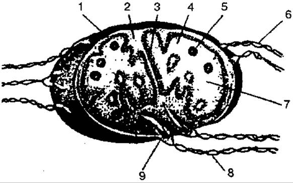
Fig. 98. Structure of a lymph node:
1 — capsule; 2 — trabecula; 3 — cord; 4 — cortex; 5 — follicles; 6 — afferent lymphatic vessels; 7 — medulla; 8 — efferent lymphatic vessels; 9 — hilum of the lymph node
Each lymph node is externally covered by a Connective Tissue capsule. On one side, the lymph node features veins and efferent lymphatic vessels. Afferent vessels approach the node from its convex side. Thin septa extend inward from the capsule, interconnecting deep within the node.
A cross-section of the node reveals a dense peripheral cortex, consisting of cortical and paracortical zones, and a central medulla. B- and T-lymphocytes are formed within the cortex and medulla, where a leucocytic factor that stimulates Cell proliferation is also produced. Mature lymphocytes enter the nodal sinuses and are then carried by the lymph into the efferent vessels.
Last update: 08/08/2026
Editorial and Educational Adaptation: This material has been compiled based on the primary/original source text. The project team performed an editorial review, corrected technical inaccuracies, structured sections, and adapted the content for an educational format.
What was processed:
- elimination of formatting defects (OCR errors, structural breaks, corrupted characters);
- editorial organization of content;
- standardization of terminology in accordance with academic sources;
- verification of factual statements against the original source text.
All mentions of the author, publication year, and origin of the primary text have been preserved in accordance with the source.