Human Anatomy Part 1 - K. A. Diubenko, A. K. Kolomiitsev, Iu. B. Chaikovsky 2002
Special Part
Splanchnology, splanchnologia [the study of viscera] - Digestive system, systema digestorium
Mouth, oral cavity - Oral cavity proper - Tongue
The Tongue, lingua (Greek glossa, whence the pathological inflammatory condition of the tongue Tissues, glossitis), is a muscular organ composed of striated Muscle fibers, externally covered by a mucous membrane containing glands and lymphoid formations (Fig. 197). The tongue performs A number of Functions: it participates in chewing, food mixing, swallowing, and perceiving the properties and quality of food. When the Oral Cavity is closed, the tongue completely fills it; its dorsum contacts the Hard and Soft palates, while its edges and tip Touch the inner surface of the alveolar processes.
The tongue is anatomically divided into the apex, apex, body, corpus, and ROOT, radix. The upper surface of the tongue is called the dorsum of the tongue, dorsum linguae. It is divided into two parts: the anterior and larger oral part, pars oralis, and the posterior pharyngeal part, pars pharyngeal, directed toward the Pharynx. Between these parts lies a small depression, the cecal foramen of the tongue, foramen coecum linguae, which is a remnant of the thyroglossal duct, ductus thyroglossalis. Along the midline of the dorsum, the median sulcus of the tongue, sulcus medianus linguae, is visible. Additionally, the terminal sulcus, sulcus terminalis, extends to the right and left from the cecal foramen.
The bulk of the tongue consists of Muscles, mm. linguales, formed by bundles of muscle fibers running in three mutually perpendicular directions:
Class="center">
Fig. 197. The tongue, lingua (superior view)
longitudinal, transverse, and vertical. Depending on their Location, the Muscles of the tongue are classified into intrinsic and extrinsic. According to their function, the tongue muscles are divided into:
- muscles that lengthen and flatten the tongue, m. genioglossus, m. verticalis;
- muscles that shorten the tongue and move it upward, m. styloglossus, m. longitudinalis superior et inferior;
- muscles that shorten the tongue and move it downward, or laterally upon unilateral action, m. hyoglossus, m. transversus linguae.
The first group of muscles originates from the derivatives of the first pharyngeal arch; the second, from the derivative of the second pharyngeal arch; and the third, from the derivative of the third pharyngeal arch.
The intrinsic group of tongue muscles includes (Figs. 198, 199):
- superior longitudinal muscle, m. longitudinalis superior;
- inferior longitudinal muscle, m. longitudinalis inferior;
- transverse muscle of the tongue, m. transversus linguae;
- vertical muscle of the tongue, m. verticalis linguae.
1. The superior longitudinal muscle, m. longitudinalis superior, is a paired muscle that originates in the region of the tongue root, extends along the entire length of the tongue, and inserts into its apex.
Function: shortens and thickens the tongue; upon unilateral contraction, deviates it to the side.

Fig. 198. Muscles of the newborn tongue in sagittal section (after Spalteholz):
1 - m. transversus linguae; 2 - m. longitudinalis inferior; 3 - m. longitudinalis superior; 4 - tunica mucosa linguae; 5 - foramen caecum; 6 - glandulae linguales; 7 - epiglottis; 8 - corpus ossis hyoidei; 9 - m. geniohyoideus; 10 - m. genioglossus
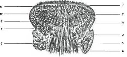
Fig. 199. Frontal section through the body of the newborn tongue (after Spalteholz):
1 - tunica mucosa linguae; 2 - m. styloglossus; 3 - septum linguae; 4 - a. profunda linguae; 5 - glandula sublingualis; 6 - m. genioglossus; 7 - n. lingualis; 8 - m. longitudinalis inferior; 9 - m. transversus linguae; 10 - m. verticalis linguae; 11 - m. longitudinalis superior
2. The inferior longitudinal muscle, m. longitudinalis inferior, is a paired muscle that originates in the region of the tongue root, passes between the m. genioglossus and m. hyoglossus, and inserts into the tongue fascia at its apex.
Function: shortens the tongue and arches the dorsum upward; upon unilateral contraction, deviates it to the side.
3. The transverse muscle of the tongue, m. transversus linguae, is a paired muscle that originates from the septum linguae and runs between the superior, inferior, and genioglossus muscles. Muscle fascicles attach to the lingual fascia on the lateral aspects of the dorsum of the tongue, while the posterior fibers reach the soft palate and pharyngeal wall.
Function: narrows and lengthens the tongue, constricts the ISTHMUS OF THE fauces and the pharynx.
4. The vertical muscle of the tongue, m. verticalis linguae, is a paired muscle originating in the region of the dorsum of the tongue from the lingual fascia and inserting into the inferior Fascia of the tongue.
Function: lengthens the tongue, flattens it, and forms a longitudinal ridge on the dorsum.
The extrinsic muscles of the tongue include (Fig. 200):
- genioglossus muscle, m. genioglossus;
- hyoglossus muscle, m. hyoglossus;
- styloglossus muscle, m. styloglossus;
- palatoglossus muscle, m. palatoglossus.
1. The genioglossus muscle, m. genioglossus, is a paired muscle that originates from the mental spine, spina mentalis, follows a spindle-shaped course, and inserts into the dorsum of the tongue and the lingual fascia.
Function: protrudes the tongue forward, and upon unilateral contraction, deviates it to the side.

Fig. 200. Muscles of the tongue - lateral view (after Spalteholz):
1 - m. styloglossus; 2 - papillae foliatae; 3 - dorsum linguae; 4 - mandibula; 5 - m. genioglossus; 6 - m. geniokyoideus; 7 - corpus ossis hyoider, 8 - m. hyoglossus; 9 - m. constrictor pharyngis medius; 10 - lig. stylohyoideum; 11 - m. stylopharyngeus
2. The hyoglossus muscle, m. hyoglossus, is a paired muscle originating from the Hyoid bone, with bundles of fibers inserting into the lingual fascia and the dorsum of the tongue.
Function: depresses and retracts the tongue, flattening it.
3. The styloglossus muscle, m. styloglossus, is a paired muscle that originates from the styloid process of the Temporal bone and the lig. stylomandibulare, directed medially and downward, blending into the lateral PARTS OF THE tongue.
Function: elevates and retracts the tongue upward and backward.
4. The palatoglossus muscle, m. palatoglossus, originates from the transverse muscle of the tongue, runs within the anterior palatoglossal arch, and extends into the palate.
Function: depresses the soft palate and constricts the isthmus of the fauces.
The mucous membrane of the tongue is externally covered by stratified squamous epithelium. It contains taste Organs, glands, and lymphoid tissue (lingual tonsil, tonsilla lingualis). In the region of the apex, dorsum, and root of the tongue, the mucous membrane is rough, and posterior to the terminal sulcus, sulcus terminalis, it features nodular elevations formed by accumulations of lymphoid tissue. The Inferior surface of the lingual mucosa is smooth and forms the frenulum of the tongue, frenulum lingue, along the midline.
The mucous membrane of the posterior part of the tongue forms three glosso-epiglottic folds: an unpaired median fold, plica glosso-epiglotica mediana, and paired lateral folds, рlicа glosso-epiglotica laterales, between which lie the epiglottic valleys, valleculae epigloticae.
On the superior and lateral surfaces of the tongue, there are mucosal elevations known as lingual papillae (see Fig. 197). The lingual papillae are divided into six types:
- filiform papillae, papillae filiformes;
- conical papillae, papillae сопісае;
- fungiform papillae, papillae funiformes;
- vallate papillae, papillae vallatae;
- foliate papillae, papillae folliatae;
- lentiform papillae, papillae lentiformes.
The most numerous are filiform papillae, which cover the entire dorsal surface of the tongue. Fungiform papillae are mainly distributed around the tip of the tongue, whereas foliate papillae are located on the lateral margins as 3 to 8 vertically oriented folds, 2 to 5 mm in length, separated by narrow vallate papillae. Vallate papillae are situated at the boundary between the root and the body of the tongue. Their arrangement forms a V-shaped line (sulcus terminalis), the apex of which points backward and reaches the foramen cecum of the tongue (a remnant of the thyroglossal duct).
The receptor apparatus of the gustatory analyzer is housed within the taste buds of the lingual papillae. It is generally accepted that fungiform and foliate papillae detect sour, sweet, and salty tastes, while vallate papillae perceive bitter tastes.
Beneath the mucous membrane lie the lingual glands, which are classified into serous, mucous, and mixed types. Their ducts predominantly open onto the inferior surface of the tongue and into the sulci surrounding the vallate papillae.
The Blood supply to the tongue is provided by the a. lingualis, while venous drainage occurs via the corresponding vein, which empties into the Internal jugular vein.
Lymphatic drainage flows into the submandibular, submental, and retropharyngeal Lymph Nodes.
The Innervation of the oral mucosa of the tongue is distributed as follows: the anterior two-thirds are innervated by the n. lingualis; the posterior third by the n. glossopharyngeus; the root region by the n. laryngeus superior (derived from the n. vagus); the taste buds of the fungiform and foliate papillae receive nerve fibers via the chorda tympani from the n. intermedius; and the vallate papillae are innervated by the n. glossopharyngeus. The intrinsic and extrinsic muscles of the tongue are innervated by the Hypoglossal nerve, n. hypoglossus (XII).
Last update: 08/08/2026
Editorial and Educational Adaptation: This material has been compiled based on the primary/original source text. The project team performed an editorial review, corrected technical inaccuracies, structured sections, and adapted the content for an educational format.
What was processed:
- elimination of formatting defects (OCR errors, structural breaks, corrupted characters);
- editorial organization of content;
- standardization of terminology in accordance with academic sources;
- verification of factual statements against the original source text.
All mentions of the author, publication year, and origin of the primary text have been preserved in accordance with the source.