Human Anatomy, Part 1 - K. A. Dyubenko, A. K. Kolomiyytsev, Yu. B. Chaykovsky 2002
Special Section
Myology, myologia [the study of muscles] — Muscles and Fascia of the Trunk
Muscles of the Back, musculi dorsi — Deep Muscles of the Back
The Deep Muscles of the back form three layers: superficial, intermediate, and deep. The superficial layer consists of three muscles: the splenius capitis, splenius cervicis, and erector spinae. The intermediate layer comprises the transversospinales muscles, while the deep layer includes the intertransversarii, interspinales, and suboccipital muscles. The superficial muscles cover vast areas of the back, sacrum, and posterior neck, reaching as far as the Occipital bone. These are powerful muscles that primarily perform static work, maintaining THE Vertebral Column in an upright position. The intermediate muscles run obliquely, spanning from the transverse processes of the vertebrae to the spinous processes. In the deep layers, the Muscle bundles are short and attach to adjacent vertebrae, whereas in the superficial layers, they are longer and span across 5-6 vertebrae. In the third (deep) layer, short muscles are located between the spinous and transverse processes of the vertebrae; they are particularly well-developed in the cervical and lumbar Regions of the vertebral column.
1. Splenius capitis muscle, m. splenius capitis.
2. Splenius cervicis muscle, m. splenius ceruicis.
3. Erector spinae muscle, m. erector spinae:
1) iliocostalis muscle, m. iliocostalis;
2) longissimus muscle, m. longissimus;
3) spinalis muscle, m. spinalis.
4. Transversospinales muscles, m. transversospinales:
1) semispinalis muscle, m. semispinalis;
2) multifidus muscles, mm. multfidi;
3) rotators, mm. rotatores.
5. Intertransverse muscles, mm. intertransuersarii.
6. Interspinal muscles, mm. interspinales.
7. Levatores costarum muscles, mm. levatores costarum.
1. Splenius capitis muscle, m. splenius capitis - located deep to the m. trapezius (Fig. 126). It originates from the nuchal ligament and the spinous processes of the 3rd cervical to 3rd thoracic vertebrae, inserting into the lateral third of the superior nuchal line and the lateral aspect of the mastoid process.
Function: bilateral contraction extends the cervical spine and the HEAD, whereas unilateral contraction rotates the head to the same side.
Blood supply: occipital and deep cervical Arteries (aa. occipitalis, cervicalis profunda).
Innervation: cervical nerves (nn. cervicales, CIV-CVII).
2. Splenius cervicis muscle, m. splenius cervicis (see Fig. 126), located in the posterior cervical region. It originates from the spinous processes of the 3rd to 5th thoracic vertebrae and inserts onto the posterior tubercles of the transverse processes of the upper 2nd to 3rd cervical vertebrae.
Class="center">
Fig. 126. Back muscles: superficial muscles — third layer; deep muscles — First and Second layers (after R. D. Sinelnikov)
Function: bilateral contraction pulls the Head and Neck backward, while unilateral contraction rotates them to the same side.
Blood supply: deep cervical and occipital arteries (aa. cervicalis profunda, occipitalis).
Innervation: greater occipital nerve, cervical nerves (n. occipitalis major, nn. cervicales, CII-CVIII).
3. Erector spinae muscle, m. erector spinae — a powerful muscle situated in the superficial layer along the entire length of the vertebral column, flanking the line of the spinous processes from the sacrum to the head (see Figs. 126, 127). It originates from the dorsal surface of the sacrum (facies dorsalis), the spinous processes of the lumbar vertebrae, the posterior part of the iliac crest (crista iliaca), and the thoracolumbar fascia (fascia lumbodorsalis); it ascends and progressively divides into three columns: lateral, intermediate, and medial.
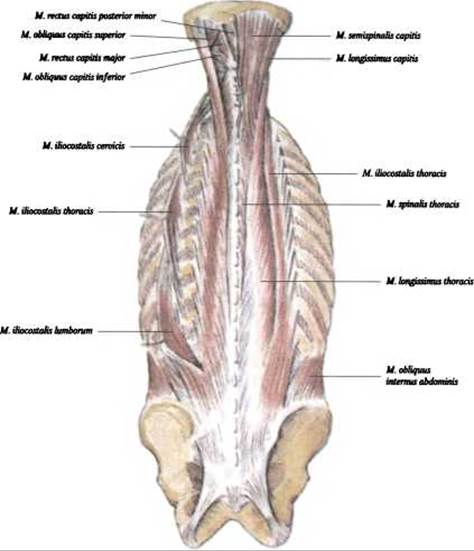
Fig. 127. Deep back muscles: superficial layer and suboccipital muscles (according to R. D. Sinelnikov)
1) Lateral tract - iliocostalis muscle, m. iliocostalis (see Fig. 127), located most laterally and divided into three parts:
- m. iliocostalis lumborum - attaches to the angles of the lower six or seven Ribs;
- m. iliocostalis thoracis - originates by separate slips from the lower six ribs and attaches to the angles of the upper six;
- m. iliocostalis cervicis - originates from the upper five or six ribs and attaches to the posterior tubercles of the transverse processes of cervical vertebrae IV-VI.
Function: extends and straightens the vertebral column, and upon unilateral
contraction, laterally flexes the vertebral column to its own side and depresses the rib.
Blood supply: aa. intercostales posteriores, aa. lumbales.
Innervation: rr. dorsales nn. spinales (CIV-IIII).
2) Intermediate tract - longissimus muscle, m. longissimus, located between m. iliocostalis and m. spinalis (see Fig. 127). It is divided into the following four
muscles:
- longissimus lumborum muscle, m. longissimus lumborum — attaches via medial fascicles to the accessory processes of the lumbar vertebrae, and via lateral fascicles to the transverse processes of these vertebrae;
- longissimus thoracis muscle, m. longissimus thoracis — receives additional fascicles from the transverse processes of the six lower thoracic vertebrae and attaches to the angles of the ten lower ribs and the posterior PARTS OF THE transverse processes of all thoracic vertebrae;
- longissimus cervicis muscle, m. longissimus cervicis — originates from the transverse processes of the upper V–VI thoracic vertebrae and attaches to the transverse processes of the II–V cervical vertebrae;
- longissimus capitis muscle, m. longissimus capitis — originates from the transverse processes of the upper three thoracic and three cervical vertebrae, and attaches to the mastoid process.
Function: the longissimus cervicis and thoracis muscles extend the Vertebral Column and laterally flex it; the longissimus capitis extends the head and turns the face toward the same side.
Blood supply: lumbar arteries, posterior intercostal arteries, deep cervical artery.
Innervation: dorsal rami, Spinal Nerves (C II–V).
3) Medial tract — spinalis muscle, m. spinalis (see Fig. 127), is divided into two muscles:
- spinalis thoracis muscle, m. spinalis thoracis — originates from the spinous processes of the two upper lumbar and two lower thoracic vertebrae, skips one or two vertebrae, and attaches to the spinous processes of the III–VIII thoracic vertebrae;
- spinalis cervicis muscle, m. spinalis cervicis — originates from the spinous processes of the upper two thoracic and lower two cervical vertebrae, and attaches to the spinous processes of the II–IV cervical vertebrae.
Function: bilateral contraction extends the spine and maintains an upright posture, while unilateral contraction laterally flexes the spine to the corresponding side; the upper fascicles of the muscle pull the head to the same side, and some fascicles depress the ribs.
Blood supply: posterior intercostal arteries, deep cervical artery.
Innervation: dorsal rami of spinal nerves (C I–C II).
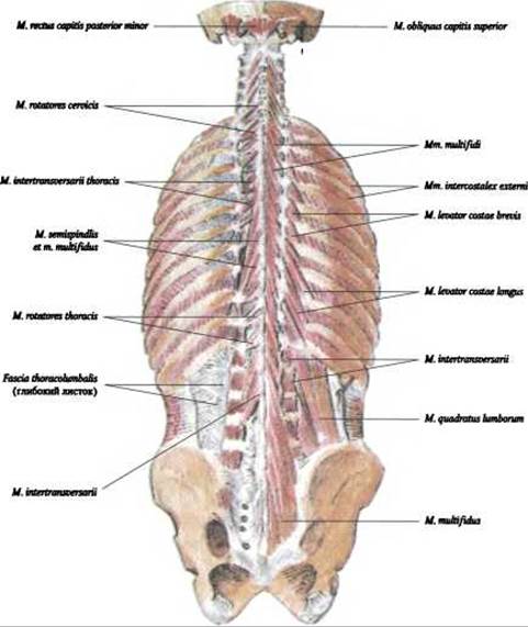
Fig. 128. Deep back muscles – second and third layers (after R. D. Sinelnikov)
4. Transversospinales muscle, m. transversospinalis — located extending from the sacrum to the occipital bone, beneath the m. longissimus and m. spinalis, filling the groove between the spinous and transverse processes. The muscle bundles have an oblique direction and span from the transverse processes of inferior vertebrae to the spinous processes of superior ones. Three muscle layers are distinguished: the superficial layer, m. semispinalis, the middle layer, mm. multifidi, and the deep layer, mm. rotatores (Fig. 128).
1) Semispinalis muscle, m. semispinalis — its bundles span across five to six vertebrae; this muscle is absent in the lumbar region. It is subdivided into the semispinalis Muscles of the Thorax, neck, and head, m. semispinalis thoracis, cervicis et semispinalis capitis. The muscle originates from the transverse processes of all thoracic and the six lower cervical vertebrae, and inserts into the spinous processes of the six lower cervical vertebrae: the semispinalis capitis muscle, m. semispinalis capitis, is located on both sides of the lig. nuchae. It originates from the transverse processes of the six upper thoracic and four lower cervical vertebrae, and inserts into the occipital bone between the superior and inferior nuchal lines.
Function: upon contraction of all bundles, the muscle extends the upper sections of the spine and pulls the head backward or holds it in an extended position; unilateral contraction causes slight rotation.
Blood supply: aa. intercostales posteriores, a. cervicalis profunda.
Innervation: r. dorsalis nn. spinales (CII—CV; ThI-ThIII).
2) Multifidus muscles, mm. multifidi (see Fig. 128) — their bundles span across three to four vertebrae, extending from the second cervical vertebra to the sacrum. They originate from the facies dorsalis of the sacrum, processus mamillares of the lumbar vertebrae, processus transversi of the thoracic vertebrae, and processus articulares of the four lower cervical vertebrae, and insert into the processus spinosi of all lumbar, thoracic, and cervical vertebrae, except for the atlas.
Function: rotate the vertebral column around its longitudinal axis, and participate in extension and lateral flexion.
Blood supply: aa. lumbales, aa. intercostales posteriores, a. cervicalis profunda.
Innervation: r. dorsalis nn. spinales (CII-SI).
3) Rotator muscles, mm. rotatores — form the deepest layer of the back musculature (see Fig. 128). They span across one vertebra or insert into the adjacent one. Long and short rotators are distinguished — mm. rotatores longi and mm. rotatores breves. The latter, located between adjacent vertebrae, connect the transverse processes of one vertebra to the Base of the spinous process of another; m. rotatores longi skip one, rarely two vertebrae.
Function: the transversospinalis muscle extends the spine upon bilateral contraction, and rotates it to the opposite side upon unilateral contraction.
Blood supply: a. cervicalis profunda, aa. intercostales posteriores, aa. lumbales.
Innervation: rr. spinales (C II-C IV).
5. Intertransverse muscles, m. intertransversarii — located in the cervical and lumbar regions (see Fig. 128). In the lumbar region, lateral muscles (between transverse processes) and medial muscles (between processus accessorii of adjacent vertebrae) are distinguished.
Function: stabilize the spine, and bend it laterally upon unilateral contraction.
Blood supply: a. cervicalis profunda, aa. intercostales posteriores, aa. lumbales.
Innervation: rr. dorsales nn. spinales (C I-C IV; L I-L IV).
6. Interspinal muscles, mm. interspinales — located between the apices of the spinous processes of adjacent vertebrae in the cervical and lumbar regions (see Fig. 128).
Function: extend the spine and maintain it in an upright posture.
Blood supply: a. cervicalis profunda, aa. intercostales posteriores, aa. lumbales.
Innervation: rr. dorsales nn. spinales (CIII-IV).
7. Levator costarum muscles, mm. levatores costarum — developed only in the thoracic region. They originate from the transverse processes of the VII cervical and I-XI thoracic vertebrae, and run laterally and inferiorly to the angle of the nearest rib. In addition to these short muscles, the four lower ribs also feature long ones that skip one rib and insert into the next.
Function: elevate the ribs.
Blood supply: aa. intercostales posteriores.
Innervation: nn. spinales, nn. intercostales (C VIII, Th I-Th XI).
Last update: 08/08/2026
Editorial and Educational Adaptation: This material has been compiled based on the primary/original source text. The project team performed an editorial review, corrected technical inaccuracies, structured sections, and adapted the content for an educational format.
What was processed:
- elimination of formatting defects (OCR errors, structural breaks, corrupted characters);
- editorial organization of content;
- standardization of terminology in accordance with academic sources;
- verification of factual statements against the original source text.
All mentions of the author, publication year, and origin of the primary text have been preserved in accordance with the source.