Human Anatomy, Part 1 - K. A. Dyubenko, A. K. Kolomiytsev, Y. B. Chaykovsky 2002
Special Part
Myology, myologia [the study of muscles] — Muscles and Fasciae of the Trunk
Muscles of the Back, musculi dorsi — Superficial Muscles of the Back
Based on their Location, the back Muscles are divided into superficial and deep. The superficial muscles attach to the Pectoral Girdle and arm (trapezius, latissimus dorsi, levator scapulae, rhomboideus) and the Ribs (serratus posterior superior and serratus posterior inferior). The deep muscles (autochthonous) are located in the grooves lateral to THE Vertebral Column, bounded by the ribs, transverse processes, and spinous processes of the vertebrae.
The superficial back muscles form two layers.
First layer:
1. Trapezius Muscle, m. trapezius.
2. Latissimus dorsi muscle, m. latissimus dorsi.
3. Levator scapulae muscle, m. levator scapulae.
Second layer:
4. Rhomboideus muscle, m. rhomboideus.
5. Serratus posterior superior muscle, m. serratus posterior superior.
6. Serratus posterior inferior muscle, m. serratus posterior inferior.
1. Trapezius muscle, m. trapezius — a flat, broad muscle occupying the upper back and occipital region (Fig. 124). It is triangular in shape, with its base aligned with the line of the spinous processes of the vertebrae and its apex lying on the acromion. Together, the right and left trapezius muscles form an irregular quadrilateral, hence the name trapezius. The contours of this muscle are clearly visible in a living person. In the region of the VII cervical vertebra, it forms a tendinous "mirror" shaped like a rhombus. The muscle originates with short tendinous bundles from the superior nuchal line, nuchal ligament, external occipital protuberance, spinous processes of the VII cervical and I–XII thoracic vertebrae, and the supraspinous ligament. Insertion: upper fibers attach to the acromial end of the clavicle; middle fibers to the acromion; lower fibers to the spine of the scapula.
Class="center">
Fig. 124. Superficial back muscles — First and Second layers (after V. P. Vorobyov)
Function: contraction of the middle bundles of the muscle draws the scapula toward the spine. Contraction of the upper bundles elevates the pectoral girdle, while the lower bundles depress it. When the scapula is fixed, both trapezius muscles pull the HEAD backward; unilateral contraction turns the head to the side opposite the contracting muscle. When the Vertebral Column and head are fixed, the upper part of this muscle elevates the scapula.
Blood supply: transverse cervical, occipital, suprascapular, and posterior intercostal Arteries.
Innervation: external branch of the Accessory nerve, cervical nerves (CII–CIV).
2. Latissimus dorsi muscle, m. latissimus dorsi — located directly beneath the Skin, covered by the trapezius muscle only in its upper part; it occupies the lower back and the lateral chest region (see Fig. 124). It is well-developed in athletes (rowers, skiers). It originates via a tendinous aponeurosis from the spinous processes of the lower V–VI thoracic vertebrae, all lumbar and sacral vertebrae, as well as the median sacral crest, the posterior third of the external lip of the iliac crest, and the lower three to four ribs. In its upper part, the muscle covers the inferior margin of the teres major muscle. It inserts via a flat tendon into the crest of the lesser tubercle of the humerus.
Function: adducts and pronates the arm, depresses the pectoral girdle, and draws the scapulae toward the vertebral column. When the upper limbs are fixed, it draws the trunk toward them.
Blood supply: thoracodorsal, posterior circumflex humeral, and posterior intercostal arteries.
Innervation: thoracodorsal nerve (CVI–CVIII).
3. Levator scapulae muscle, m. levator scapulae — located in the lateral Regions of the posterior neck, beneath the trapezius muscle (see Fig. 124). It originates from the tubercles of the transverse processes of the upper four cervical vertebrae. It inserts into the medial angle and part of the medial border of the scapula.
Function: when the vertebral column is fixed, it elevates the scapula; when the scapula is fixed, acting together with the Muscles of the opposite side (sternocleidomastoid and scalene), it flexes the cervical spine toward the contracting side, while bilateral contraction maintains the neck in a vertical position.
Blood supply: transverse cervical, superficial cervical, and ascending cervical arteries.
Innervation: dorsal scapular nerve (CIII–CV).
4. Rhomboideus muscle, m. rhomboideus — located beneath the trapezius muscle and rhomboid in shape (see Figs. 124, 125). It originates from the spinous processes of the lower two cervical and upper four thoracic vertebrae, and inserts into the medial border of the scapula.
Function: adducts the scapula toward the median plane of the trunk and elevates it.
Blood supply: aa. transversa colli, suprascapularis, intercostales posteriores.
Innervation: n. dorsalis scapulae (CIV-CV).
5. Serratus posterior superior muscle, m. serratus posterior superior — a thin muscle located beneath the m. rhomboideus (see Fig. 125). It originates from the lower part of the nuchal ligament and the spinous processes of the lower two cervical and upper two thoracic vertebrae. It attaches by four slips to the upper ribs, from II to V, lateral to their angles.
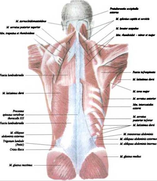
Fig. 125. SUPERFICIAL MUSCLES OF the back — second and third layers (after V. P. Vorobyov)
Function: elevates the upper ribs and participates in inspiration.
Blood supply: aa. intercostales, cervicalis profunda.
Innervation: nn. intercostales (ThI-ThIV).
6. Serratus posterior inferior muscle, m. serratus posterior inferior — located beneath the m. latissimus dorsi (see Fig. 125). It originates from the spinous processes of the lower two thoracic and upper two lumbar vertebrae. It attaches by four slips to the four lower ribs.
Function: depresses the lower ribs and participates in inspiration.
Blood supply: aa. intercostales posteriores.
Innervation: nn. intercostales (ThX-ThXII).
Last update: 08/08/2026
Editorial and Educational Adaptation: This material has been compiled based on the primary/original source text. The project team performed an editorial review, corrected technical inaccuracies, structured sections, and adapted the content for an educational format.
What was processed:
- elimination of formatting defects (OCR errors, structural breaks, corrupted characters);
- editorial organization of content;
- standardization of terminology in accordance with academic sources;
- verification of factual statements against the original source text.
All mentions of the author, publication year, and origin of the primary text have been preserved in accordance with the source.