Antibiotics (Properties, Application, Interactions) - M.P. Cherenko 1999
Anaerobic Infection (Gas Gangrene)
CLOSTRIDIAL (SPORE-FORMING) ANAEROBIC INFECTION
Anaerobic clostridial infection, commonly known by the synonym "gas gangrene," is, much like tetanus (another specific anaerobic infection), a wound toxemia. It typically accompanies combat injuries. During the Great Patriotic War, it was relatively rare—affecting 0.5–1% of the wounded (though in previous conflicts it reached near-epidemic proportions, with complications recorded in 5–13% of wounded personnel During the first World War). However, despite its relative rarity, gas gangrene still occurs in peacetime, particularly following railway and automobile accidents, domestic and street knife wounds penetrating the intestine, and so forth. Occasionally, it develops postoperatively (lower limb amputations due to gangrene, digestive tract surgeries including colorectal Procedures, post-abortion complications—especially illegal abortions—and even following injections). The causative agents of this infection are anaerobic clostridia, primarily Cl. perfringens (a gas-producing clostridium), Cl. oedematiens (an edema-forming clostridium), Cl. histolyticum (a tissue-dissolving clostridium), and Cl. septicum. Other microorganisms (clostridia) do not play an independent role in the Pathogenesis of the disease.
Anaerobic microflora inhabit the Large Intestine of animals and humans, from where they enter the environment (via feces, upon the death of animals and humans) into soil and air, where they can persist for long periods, particularly in spore form.
Garden plots, orchards, and strips along railway lines fertilized with animal and human manure (organic fertilizers) are particularly rich in clostridia.
Clostridia are detected in such soils even at considerable depths (up to 1 m). This explains why anaerobic infection was more frequently observed during wartime defensive operations, when troops remained stationary for prolonged periods in terrain contaminated with feces. Cl. perfringens is present in the digestive tract of 25–30% of healthy individuals and in the Urogenital System of many adult women. It is detected on the Skin of up to 44% of uninfected patients treated in hospitals (N. M. Sommers, 1980), as well as in the urogenital tracts of women. Under such circumstances, patients face a potential risk of developing gas gangrene under favorable conditions. Nevertheless, the low prevalence of clostridia remains a decisive factor in The Development of the infection.
The primary factor in the pathogenesis of the condition is the presence of favorable conditions for clostridia or their spores to thrive, namely: unventilated wound pockets and necrotic Tissues, particularly Muscles; and an increased susceptibility of the body to these pathogens due to various causes—impaired general Circulation, Metabolic Disorders (Diabetes Mellitus, hypovitaminosis, hypoproteinemia, etc.), anemia, chilling, and immune suppression. This is evidenced by the fact that while contamination of injuries (such as railway trauma, which are typically open) is recorded in virtually all cases, the infection develops in only about 1% of victims. The Role of delayed surgical wound debridement in the pathogenesis of anaerobic infection is particularly noteworthy.
Anaerobic infection most commonly develops in deep penetrating gunshot, lacerated, contused, and stab wounds of the lower extremities (leg and thigh — 75%), buttocks, and trunk. Among other locations, it most frequently complicates wounds of the upper extremities and abdomen, less frequently the chest, and least frequently the neck and HEAD (due to the rich vascularization of the latter). The high frequency of anaerobic infection in wounds of the lower limbs and buttocks is explained both by the presence of large Muscle masses in these areas (with high Glycogen and protein content) and by their heavy contamination with clostridia due to their proximity to the anus and urogenital Organs.
Wounds of the extremities complicated by bone fractures or major vascular injury (and their ligation) are particularly dangerous, as bone fragments cause muscle damage and tissue necrosis, while vascular trauma leads to tissue ischemia and necrosis. In gunshot wounds, the role of bone damage (fractures) in the onset of wound infection is especially striking (the infection is observed several times more frequently with fractures than without them). Anaerobic infection develops more readily in blind wounds, especially splinter wounds, because splinters carry soil, clothing fragments, and other vehicles of implantational infection while causing more extensive tissue necrosis. When introduced with soil, calcium and silicon also promote the Development of the infection.
Anaerobic infection is chiefly caused by associations of anaerobic clostridia, and less frequently by a monoculture. The most common combination is the gas-producing Cl. perfringens, isolated in 56–100% of patients, and the edema-producing Cl. oedematiens (in 15–50% of patients), while combinations of three clostridia are rarer (A. N. Lvov, O. A. Dolina, 1964; W. A. Altemeier, 1995). Cl. histolyticum is encountered least often. Along with anaerobic clostridia and non-spore-forming anaerobes, associations frequently harbor pathogens of pyogenic (banal) acute infections, specifically staphylococci and streptococci. By consuming oxygen, the latter create favorable conditions for clostridial growth and enhance their virulence.
Classification. The clinical diversity of anaerobic infection variants hinders the establishment of a universally accepted single classification. Numerous terms are used to designate it: gas infection, gas gangrene, anaerobic gangrene, acute malignant edema, bronze erysipelas, white erysipelas, etc. The terms "anaerobic infection" and "gas gangrene" are most widely used.
There are numerous, predominantly clinical, classifications of this infection. Among them, the most commonly used is the classification proposed by M. Weinberg and P. Séguin (1918), which identifies 5 forms of infection (based on the two Main Properties of clostridia—gas production and edema): 1) emphysematous (classical); 2) edematous (toxic); 3) mixed; 4) putrefactive; 5) phlegmonous.
A classification close to this, yet somewhat simplified, was recently proposed by O. M. Berkutov, who distinguishes 3 forms: 1) gas; 2) gas-edematous; 3) purulent-putrefactive.
Even N. I. Pirogov distinguished the deep, subfascial form, which is primary, and the superficial, epifascial form (about 6%). This division remains in use today.
Deep forms (according to O. M. Berkutov's classification) include the gas and gas-edematous forms, whereas the purulent-putrefactive form is classified as superficial. Many foreign researchers refer to the deep forms as anaerobic myonecrosis.
Based on their clinical course, anaerobic infections are classified as fulminant, rapidly spreading, and slowly spreading.
Comprehensive classification CHARACTERISTICS OF ANAEROBIC infection forms must reflect both clinical and pathomorphological Features of the disease.
Pathological Anatomy. Upon entering conditions favorable for their development, anaerobic clostridial spores germinate into vegetative forms within the wound, releasing potent toxins (necrotoxin, hemolysin, etc.) and toxigenic Enzymes (lecithinase or alpha-toxin, hyaluronidase, collagenase, proteases).
By causing lysis of Cell membranes (including erythrocytes), toxins and enzymes increase cellular and vascular permeability, induce tissue edema and necrosis, and pave the way for microbes to colonize and multiply within the dead tissues. The infection affects muscles, Connective Tissue, Blood Vessels, and nerves. It remains uncertain which Structure is affected primarily—the muscles (myonecrosis) or the blood vessels (thrombosis resulting in myonecrosis).
Due to the utilization and Fermentation of glycogen and other CARBOHYDRATES in affected tissues, lactic acid and gases—methane, hydrogen, and CO2—accumulate, accompanied by ammonia and hydrogen sulfide if a Putrefactive infection is superadded. The tissues become edematous. The edema spreads rapidly in a central (proximal) direction, causing pallor of the skin. Frequently, blisters containing serosanguineous fluid form on the skin (due to a sharp increase in vascular permeability and accumulation of transudate beneath the epidermis). For objective recording of edema during the Great Patriotic War, it was proposed to tie a thread around the limb above the wound, which would sink deeper into the skin as the edema progressed. Gas in the tissues and tissue edema are invariably present, though in varying combinations—either gas production predominates (monoculture of Cl. perfringens) or edema does (association of Cl. perfringens with Cl. oedematiens). The necrotic process dominates in and around the wound, whereas inflammatory-reactive cellular responses are poorly expressed.
In the blood vessels (Veins and Arteries) of tissues surrounding the focus of infection (the wound), stasis and thrombosis develop (as a consequence of edema and toxin action), accompanied by myonecrosis, tissue Hemorrhage, and hemolysis. The latter give rise to map-like purplish-blue and brown patches on the skin. The wound itself turns gray, its discharge is scanty, serosanguineous or dark brown, and foul-smelling, and the muscles appear dull and protrude into the wound. Only in purulent-putrefactive epifascial forms are inflammatory reactions noticeable in the form of leukocytic infiltration in the tissues surrounding the focus of infection (wound).
In the presence or predominance of Cl. histolyticum within the microbial association, all tissues undergo lysis, turning into a liquid mixture enclosed within the skin.
Clinical Course. Signs and Symptoms. In the vast majority of patients, the disease occurs in lower extremity injuries (thigh, leg), developing chiefly during the first week post-injury—most often within the first 3 days, frequently prior to the development of pyogenic infection. The belief that the presence of the latter rules out the possibility of an anaerobic infection is mistaken. The shorter the incubation period, the more severe the clinical course of the disease.
The initial local signs are a sensation of dull, bursting pain in the wound, a feeling of tightness from the bandage if the wound is located on a limb, decreased wound exudation, A change in wound color from pinkish-red to gray, and the appearance of edema in surrounding tissues, particularly proximal to the wound. General manifestations in the early period include a feeling of weakness, anxiety, agitation (followed later by depression), and marked tachycardia disproportionate to the body Temperature. The temperature is most often subfebrile, and less frequently febrile. Local changes follow a phased course, progressing from initial signs—a localized gas Phlegmon (early phase)—through gas phlegmon to gangrene.
In cases of delayed medical care—such as untimely surgical intervention, including wound debridement or amputation, or in uncontrolled disease progression—the spread of necrotic changes leads to microbial invasion of the bloodstream, the development of anaerobic Sepsis, and patient death.
Class="center">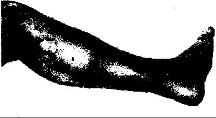
Fig. 76. Gas Gangrene of the lower extremity
In the stage of anaerobic phlegmon—both localized and, to an even greater extent, widespread—the wound becomes grayish-muddy, dry (in gas gangrene) or more moist (in the gas-necrotic form), with a small amount of serosanguinous or turbid, foul-smelling exudate. The muscles within the wound lose their luster, turn gray or hemorrhagic, and bulge into the wound. The tissues surrounding the wound are edematous. Due to vascular compression by the edema, the skin far beyond the focus becomes pale, with noticeable bluish venous streaks on its surface. Palpation of the edematous tissues around the wound reveals crepitus.
As the infection spreads through the tissues, myonecrosis, muscle necrosis (of individual muscles or muscle groups), and gangrene develop. Muscle and vascular necrosis lead to hemorrhages, Hemoglobin breakdown, and the appearance of blisters and scattered map-like patches on the skin of the affected areas of the limbs or body, exhibiting purplish-blue, brownish-yellow (bronze), and sometimes yellowish-green hues (known as "ecchymosis blooming"). Because of this, the skin discoloration is often referred to as "autumn foliage coloration."
When anaerobic infection predominantly localizes in the legs, myonecrosis spreads rapidly, causing Thrombosis of the main arteries and gangrene of one or, rarely, both legs. The pulse disappears in the Arteries of the leg (distally from the wound), and the limb becomes cold and dark blue or even bluish-black (Fig. 76).
In gangrene, the wound becomes dark, muddy, necrotic, and foul-smelling. The General condition of the patient, which is already significantly impaired during the phlegmon stage (emotional depression, low-grade fever, tachycardia, loss of appetite, dryness of the mucous membranes and Tongue, coating, etc.), deteriorates sharply with the development of widespread phlegmon, and especially myonecrosis and gangrene. Body temperature rises to febrile levels (38–40 °C), accompanied by pronounced intoxication (anorexia, depression, scleral icterus, a dry tongue coated with gray fur, dyspeptic symptoms, tachycardia, low blood pressure, leukocytosis, low urine output, progressive anemia, and apathy).
The onset of sepsis is characterized by deepening systemic symptoms of intoxication and the development of toxic Shock.
The clinical course of the disease can be fulminant, rapid, or slow, and its severity ranges from very severe and severe to moderate.
In the vast majority of cases, anaerobic infection presents as a severe myonecrotic form. Only in a small number of patients is the infection limited to the involvement of the suprafascial (epifascial) soft tissues and connective tissue proper (subcutaneous tissue, blood vessels, superficial fascia), which corresponds to the development of anaerobic phlegmon or, in the terminology of some Western authors, anaerobic cellulitis (with or without gas production). Unlike myonecrotic forms, which are predominantly characterized by a necrotic process, this form proceeds with clear signs of inflammation as well—leukocytic infiltration of the tissues around the focus accompanied by phagocytosis. The course of this form of infection is considerably milder, and the prognosis for patients is favorable.
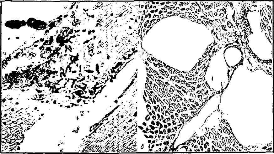
Fig. 77. Microscopy of affected muscles in gas gangrene: a — numerous gas-producing clostridial bacilli and muscle necrosis; b — gas accumulation between muscle fibers (W. Anderson)
The Diagnosis of anaerobic infection is based primarily on the clinical manifestations of the disease, including local signs and general systemic disturbances. Local signs include alterations in the appearance of the wound itself, edema of the tissues surrounding it and extending beyond it (particularly proximally when the main localization is the extremities, especially the legs), crepitus in the tissues around the focus, and scanty, foul-smelling, brownish-muddy wound discharge (in the absence of putrefactive Microorganisms in the flora, the odor is unpleasant, resembling fermented cabbage or stale cheese). Frequently, muscles bulge from the wound, resembling boiled meat or exhibiting a dark hemorrhagic color, while purplish-blue and "bronze" map-like patches and blisters appear on the skin. Radiography reveals gas in the tissues (a honeycomb, spongy STRUCTURE OF THE cellular tissue and gas accumulation in the muscles; Fig. 77; sometimes resembling a Christmas tree appearance).
Severe intoxication, which early on triggers systemic disorders—such as progressive decompensation of The Cardiovascular system and Internal Organs—along with progressive anemia, completes the clinical picture of the disease.
MICROBIOLOGICAL EXAMINATION OF wound discharge and necrotic tissues helps establish a presumptive diagnosis (the presence of large, thick, gram-positive rods with rounded ends (Cl. perfringens) and comma-shaped rods (in the presence of Cl. vibrio septicum), whereas a pure culture can only be obtained after 2 days, which is too late. Therefore, Treatment strategy is based on clinical signs and symptoms. Determining the enzymes hyaluronidase and lecithinase for diagnostic purposes—as indicators of the vital activity of Cl. perfringens and Cl. vibrio septicum—has not gained widespread acceptance due to The complexity of the assay and the indirect nature of these markers.
The treatment of anaerobic clostridial infection involves both surgical and Conservative Methods. The decisive role in the treatment regimen belongs to timely surgical debridement of necrotic tissues within healthy tissue boundaries. This is achieved by making wide incisions through the soft tissues down to healthy tissue, both superficially and in depth, and excising all devitalized tissue, primarily muscles. In cases of phlegmon or isolated Necrosis of a part of the muscle with preserved pulse in the distal part of the leg or arm, this can be accomplished without amputating the limb; however, in gangrene with loss of the pulse at the periphery of the extremity, amputation far proximal to the affected tissues is the only option (for gangrene of the lower leg, amputation of the thigh in its upper or, less commonly, middle third), often supplemented by longitudinal incision of the thigh fascia along the stump. Amputations are performed by cutting through tissues at a single level—namely, guillotine amputations rather than cone-shaped or plastic ones. Wounds are left unclosed. Drains are inserted, and the wounds are continuously irrigated with oxidizing agents (hydrogen peroxide or potassium permanganate solution).
Of great importance in treating patients with anaerobic infection is The Use of Hyperbaric Oxygenation in specialized chambers (hyperbaric chambers), introduced by Brummelcamp and Boerema (1961).
However, hyperbaric oxygenation (treatment in chambers with elevated oxygen pressure) cannot replace surgery; it is effective only in combination with the latter—wide exposure of the focus and excision of dead tissue in the same manner as an amputation.
When anaerobic infection is localized outside the limbs, wide incisions are made to undermine the skin in the affected area and excise necrotic muscles down to healthy tissue, after which the wound is irrigated with oxidizing agents and left open. Subsequently, the patient undergoes hyperbaric oxygenation in a specialized chamber.
Surgical intervention is combined with the administration of 500,000 to 1,000,000 IU of sodium benzylpenicillin every 4 hours, or synthetic Penicillins, along with detoxification solutions (polyvinylpyrrolidone), protein preparations (albumin and protein), as well as fresh blood or whole plasma.
Other important measures include corticosteroids, proteolysis inhibitors, anticoagulants, cardiovascular drugs, and supportive agents. Treatment must be administered against the Background of a high-calorie (supermetabolic) diet with a high vitamin content, delivered both enterally (including via a feeding tube) and, predominantly, parenterally. The mortality rate in anaerobic infection is high, reaching 20–50% or more. It must be remembered that this infection is prone to recurrence. Late recurrences are observed following trauma or surgery involving the removal of foreign bodies. Therefore, the disease process must be closely monitored until it completely resolves, particularly until the wound has fully healed.
Patients recover slowly. They require rehabilitation in sanatorium-resort settings.
Prophylaxis. The decisive factor in preventing anaerobic gangrene is timely, early surgical wound debridement performed with proper technique (involving wide wound exposure and excision of traumatized tissues).
Patients with wounds in whom anaerobic infection may develop postoperatively (or even before surgery) are prescribed prophylactic therapy with penicillin-group Antibiotics.
The experience of wars, particularly World War II, demonstrates that anaerobic infection develops primarily As a result of delayed or inadequate primary surgical wound debridement. All other factors, such as the absence or poor immobilization of fractures, shock, hemorrhage, etc., although contributing to the development of anaerobic infection, are of secondary importance compared to primary surgical debridement.
Prophylactic serum vaccination against anaerobic clostridia (triple vaccine) has not proven effective, nor has the Treatment of the disease with antigangrene sera, although it was practiced and advocated by some surgeons during and for some time after the war.
Moreover, this prophylaxis and therapy frequently resulted in anaphylactic shock complications. Currently, prophylaxis and treatment with antigangrene sera are no longer used.
The Prevention of anaerobic infection in the Surgical treatment of wounds, including postoperative ones where anaerobic microflora proliferates, must involve ensuring adequate tissue aeration (in some cases leaving wounds without primary closure, drainage and irrigation with oxidizing agents, administration of antibiotics, particularly benzylpenicillin sodium salt, etc.).
To prevent the nosocomial spread of anaerobic infection, such patients must be isolated from others, especially those with surgical wounds.
Medical personnel in contact with these patients must strictly observe the rules of asepsis and personal hygiene. Doctors and nurses performing surgery or changing dressings on a patient must change their gowns and gloves upon completion of the Procedure, and thoroughly wash and disinfect their hands.
Dressings and drains soaked in wound discharge must be collected in a separate container and incinerated. Patient linens and the gowns of doctors and nurses are collected separately from other laundry, transported in dedicated containers to disinfection chambers where they are disinfected after soaking in a sodium bicarbonate solution. Transport splints are also disinfected in steam chambers at a temperature of 110–120 °C. Instruments (after soaking in a 1% sodium bicarbonate solution and washing) are sterilized separately in dry-heat sterilizers.
Operating rooms and dressing rooms where surgeries or dressings are performed must be thoroughly cleaned after the procedures are completed (washing floors and walls with disinfectant solutions, airing out, and irradiating with germicidal lamps). The ward floors are cleaned and washed daily, and upon completion of the patient's treatment, the beds, furniture, walls, and air undergo complete disinfection.
Last update: 08/08/2026
Editorial and Educational Adaptation: This material has been compiled based on the primary/original source text. The project team performed an editorial review, corrected technical inaccuracies, structured sections, and adapted the content for an educational format.
What was processed:
- elimination of formatting defects (OCR errors, structural breaks, corrupted characters);
- editorial organization of content;
- standardization of terminology in accordance with academic sources;
- verification of factual statements against the original source text.
All mentions of the author, publication year, and origin of the primary text have been preserved in accordance with the source.