BOTANY WITH THE BASICS OF HYDROBOTANY (AQUATIC PLANTS OF UKRAINE) - B.Ye. Yakubenko - 2011
VI. SYSTEM OF THE PLANT WORLD
LOWER SPORE PLANTS
GENERAL CHARACTERISTICS AND FEATURES
Order Charales - Charales
It encompasses macroscopic Algae (up to 20-30-100 cm long, comprising about 150 species of the modern global flora and over 1000 fossil species) with an articulated, whorled Structure differentiated into nodes and internodes, attached to the substrate by rhizoids. They feature a highly specialized oogamous sexual process, multicellular FEMALE REPRODUCTIVE Organs (antheridia and oogonia), while motile stages are represented exclusively by spermatozoa. In general morphological appearance, they resemble the sporophytes of horsetails and grow in fresh and brackish waters (continental standing Water bodies and estuarine lagoons). The most widespread genera are Chara, Nitella, and Tolypella, which are distinguished by the presence of a "stem cortex" (absent in the latter two genera) and The structure of the coronula—single-tiered and 5-merous in Chara, versus double-tiered and 10-merous in the others.
Charophyte algae frequently form dense beds where they develop, playing a significant role in the trophic and production regime of aquatic ecosystems.
Oospores (zygotes in a resting state) serve as a food source for waterfowl, especially during autumn Migrations. From a theoretical perspective, their suitability as convenient models for physiological and biophysical research is particularly important, allowing The Study of cytoplasmic streaming and electrophysiological phenomena (in particular, potential differences and cytoplasmic resistance) using these plants. The observed excitation wave propagating along the entire Cell upon electrical stimulation indicates the presence of an Action Potential, which led to its Classification as a Nerve Impulse analogous to that of Chordates. Fossil charophytes are used in geology to determine the age of sedimentary rocks (their remains are known from the Silurian—approximately 420 million years ago).
Genus Chara - Chara (Fig. 45). The sporophytes look like a small "bush" formed by a main axis consisting of combined upright supporting threads of unlimited growth ("stems") and lateral, whorled assimilation threads of limited growth ("leaves" differentiated into nodes and internodes), as well as multicellular thread-like rhizoids anchoring the plant to the sediment. Internodes are a combination of large, elongated apical-growth Cells covered with a "cortex" (which is absent in another genus of charophytes, Nitella). Nodes and internodes are formed As a result of the division of an apical cell that undergoes double differentiation. Stem basal nodes give rise to "cortex" and "stipules"; cells from each such node form two threads, one growing upward and the other downward, whose union in the central part of the internode forms the outer protective layer of the axis. Multicellular reproductive organs develop from the Cells of the leaf nodes: the male antheridium and the female oogonium. Peripheral cells of the lower node of the main axis give rise to rhizoids. Brood buds (bulbils) form on the rhizoidal nodes, facilitating Vegetative Reproduction. The Cell wall is cellulosic, typically encrusted with calcium carbonate. The Cell contains a large vacuole with cell sap, while the Cytoplasm with Organelles has a peripheral arrangement and is divided into two zones—stable and labile.
The stable zone contains a single-layered arrangement of small disk-shaped METABOLISM/14.html">Chloroplasts lacking pyrenoids and is adjacent to the Plasmalemma, whereas the labile zone contains numerous nuclei, Mitochondria, elements of The Endoplasmic reticulum, and Myosin microfibrils attached to them. Between these zones lie parallel bundles of Actin that direct the movement of myosin microfibrils with their attached organelles. Cytoplasmic streaming at speeds up to 100 µm/s facilitates the active displacement of labile zone components along the cell axis. Internode cells in the adult state are multinucleated and divide mitotically via amitosis, whereas nodal cells are uninucleated and divide through open mitosis.
Cytokinesis is of the phragmoplast type, with the Formation of primary pores and plasmodesmata in the transverse walls between daughter cells.
Class="center">
Fig. 45. General view of Chara fragilis and a thallus fragment with multicellular reproductive organs—antheridium and oogonium
Asexual reproduction is absent. Vegetative reproduction occurs via brood buds (bulbils). The sexual process is oogamous. Reproductive organs are usually located side by side on secondary lateral "branches," with the antheridia directed downward and the oogonium upward (Fig. 46). The oogonium is represented by a single large egg cell and small accessory cells connecting it to a specialized internodal cell of the sporophyte—the stalk cell. It is enclosed by five cortical cells extending from the node located beneath the stalk cell, forming closing cells at the apex of the oogonium that constitute a single-tiered, five-celled coronula. The gametophyte complex, its cortical cells, and the coronula together make up the oogonium.
Antheridia (octants) consist of a shield (scutum), a manubrium, and spermatogenous threads (Fig. 46). They develop from the apical cell of a lateral branch, which cuts off one or two disk-shaped cells whose transverse and longitudinal divisions yield 8 octant cells. Eight peripheral cells undergo planar growth to form shields that connect laterally to constitute the wall of the antheridium, forming a spherical structure. A handle cell (manubrium) extends from each shield into the sphere's cavity, terminating in one primary and 6 secondary HEAD cells bearing long, basally bifurcated spermatogenous threads. The latter consist of 100-200 disk-shaped cells, each producing a single coiled, biflagellate spermatozoid with an asymmetric flagellar "ROOT" system covered by submicroscopic scales (Fig. 46).
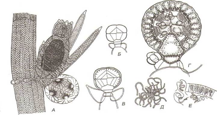
Fig. 46. General view of the reproductive organs of Chara species and their structural features:
A - reproductive organs on a "leaf"; B, C, D - cross-section of the antheridium at various developmental stages; E - manubrium (stalk),
Primary and secondary heads with spermatogenous threads; F - section of a spermatogenous thread and spermatozoa
Upon maturation of the antheridium, the mature shields separate from one another, the walls of the spermatogenous threads gelatinize, and the spermatozoa are released into the water. The cortical threads of the oogonium part beneath the coronula, creating an opening through which spermatozoa penetrate to reach the egg cell. As a result of copulation, a zygote-oospore is formed with a ribbed silica-calcareous wall, which after a resting period,
undergoes meiotic division to produce 4 haploid nuclei, three of which degenerate. Following a subsequent mitotic division, the cells differentiate, with one becoming the apical cell of a new ascending "stem"—the protonema—and the other the apical cell of the rhizoids. The life cycle is haplobiontic (haplontic), with a heteromorphic Morphology/12.html">ALTERNATION OF GENERATIONS and Sexual Dimorphism.
Last update: 07/08/2026
Editorial and Educational Adaptation: This material has been compiled based on the primary/original source text. The project team performed an editorial review, corrected technical inaccuracies, structured sections, and adapted the content for an educational format.
What was processed:
- elimination of formatting defects (OCR errors, structural breaks, corrupted characters);
- editorial organization of content;
- standardization of terminology in accordance with academic sources;
- verification of factual statements against the original source text.
All mentions of the author, publication year, and origin of the primary text have been preserved in accordance with the source.