BOTANY WITH BASICS OF HYDROBOTANY (AQUATIC PLANTS OF UKRAINE) - B.Ye. Yakubenko - 2011
VIII. BASICS OF PLANT MORPHOLOGY AND ANATOMY
Morphology and Anatomy of the Stem and Shoot
Anatomical Structure of the Stem in Monocotyledonous Plants
Monocotyledonous plants are a diverse and numerous group comprising both herbaceous and woody species that grow across a wide range of climatic and ecological conditions, resulting in considerable variation in their stem Anatomical Structure.
A typical representative of monocots with a solid stem is maize (corn) (Fig. 69). Its stem consists of the epidermis, primary cortex (during Cytology/cytology/16.html">Early stages of Organogenesis), and the central cylinder, displaying a clearly defined collateral bundle structure. The epidermis is single-layered, formed by parenchymatous Cells with thickened and lignified walls, and is covered with a thin cuticle film. Gas exchange and communication with the external environment occur through sparse Stomata. The primary cortex has a simplified structure: collenchyma is absent, and the parenchyma is represented by a thin layer of chlorophyll-bearing parenchyma, which is typically noticeable only at a young age.
Class="center">Water/water.files/image070.jpg" width="448"/>
Fig. 69. Anatomical structure of the maize stem:
1 - cuticle, 2 - epidermis, 3 - sclerenchyma, 4 - parenchymatous cells, 5 - vascular bundle, 6 - primary phloem,
7 - primary xylem, 8 - sclerenchymatous ring of the vascular bundle.
The central cylinder begins with a robust layer of sclerenchyma composed of polygonal prosenchymatous cells with uniformly thickened and lignified walls. During ontogeny, this sclerenchyma originates from pericycle cells. Its primary function is to provide structural strength to the stem, particularly against bending stresses.
Further inward lies a well-developed region of the ground parenchyma, composed of living, rounded, thin-walled cells. Closed collateral vascular bundles are scattered irregularly throughout the ground parenchyma. They consist of primary phloem—comprising sieve tubes, companion cells, and isolated phloem parenchyma cells—and xylem, which adjoins the phloem from below and contains several vessel types. Large, rounded pitted vessels are located in the central part of the xylem, interconnected by thick-walled xylem parenchyma. From this parenchyma, two smaller, rounded vessels extend toward the center: an annular-spiral vessel and an annular vessel.
These adjoin a large tear cavity formed in place of ruptured vessels. Living, thin-walled xylem parenchyma containing nutrient reserves abuts this cavity on both sides. Thin-walled xylem parenchyma is also located at the lower part of the vascular bundle, completing The formation of the closed collateral vascular bundle. It is surrounded on all sides by a robust sclerenchymatous sheath that provides mechanical protection, which is why the entire unit is referred to as a fibrovascular bundle. These vascular bundles are distributed irregularly along the perimeter of the stem.
The stem of the common bulrush (Scirpus lacustris L.) is typical of plants whose lower parts are submerged in water (Fig. 70). A dense, narrow primary cortex is covered by an epidermis containing groups of small bast fibers interspersed among its cells. Palisade chlorophyll-bearing parenchyma is located within the primary cortex. The bulk of the stem is filled with air-bearing parenchyma—aerenchyma—which forms a lattice-like network of polyhedral cells. These cells interconnect in regular chains that converge at nodes containing closed collateral vascular bundles surrounded by sclerenchymatous bundle sheaths. Diaphragms—thin plates oriented transversely across the air channels—frequently occur within the large air spaces.
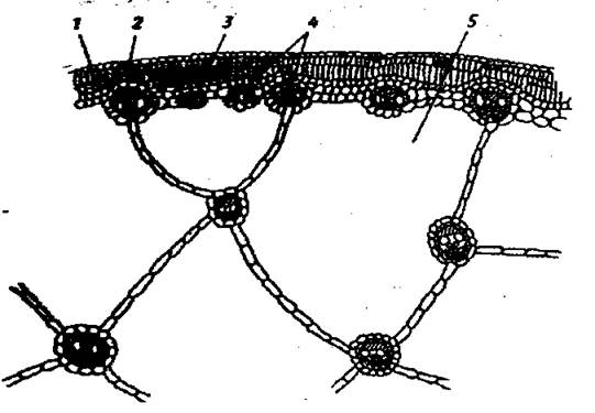
Fig. 70. Anatomical STRUCTURE OF THE common bulrush stem:
1 - epidermis, 2 - mechanical fiber bundles in the epidermis, 3 - chlorophyll-bearing parenchyma, 4 - vascular bundles, 5 - pith cells forming the aerenchyma.
The central part of the stem in the soft rush (Juncus effusus L.) is occupied by the pith, which features prominent intercellular spaces and also Functions as an aerating tissue (Fig. 71).

Fig. 71. Anatomical structure of the soft rush stem:
1 - epidermis, 2 - chlorophyll-bearing parenchyma, 3 - vascular bundle, 4 - sclerenchyma,
5 - groups of thin-walled compressed cells, 6 - stellate Cells of the aerenchyma, 7 - intercellular cavities.
These intercellular spaces are formed due to the specific Morphology of the pith cells, which possess hollow, star-shaped extensions separated by extensive air passages. The cells of this aerenchyma lack living protoplasts at maturity. Sclerenchyma is distributed in isolated patches near the stem surface, abutting the epidermis, with some portions enclosing vascular bundles and others occurring independently.
Bands of palisade chlorophyll-bearing parenchyma lie between the sclerenchyma patches, providing flexibility to the stem orientation. The vascular bundles are closed and collateral, yet they vary in size. Large bundles are enclosed by a thick sclerenchymatous layer, whereas smaller bundles feature much thinner sheaths, accompanied by secretory tissue areas whose cells are filled with mucilage.
Canadian waterweed (Elodea canadensis) belongs to helophytic plants, meaning its body is almost entirely submerged in water. The stem of Elodea is covered by a single-layered epidermis lacking a cuticle; the primary cortex has a simplified structure consisting of modified primary cortical parenchyma, while collenchyma and starch sheaths are absent. Directly beneath the epidermis lie 2–3 layers of closely packed, elongated cells rich in METABOLISM/14.html">Chloroplasts that carry out Photosynthesis—the palisade parenchyma. Deeper inward is a thick layer of aerenchyma (spongy parenchyma), which consists of parenchymatous cells linked in chains to form large air cavities. The central part of the stem is occupied by a compact central cylinder.
Last update: 07/08/2026
Editorial and Educational Adaptation: This material has been compiled based on the primary/original source text. The project team performed an editorial review, corrected technical inaccuracies, structured sections, and adapted the content for an educational format.
What was processed:
- elimination of formatting defects (OCR errors, structural breaks, corrupted characters);
- editorial organization of content;
- standardization of terminology in accordance with academic sources;
- verification of factual statements against the original source text.
All mentions of the author, publication year, and origin of the primary text have been preserved in accordance with the source.