MEDICAL BOTANY - A.G. Serbin - 2003
SECTION 1. ANATOMY
VEGETATIVE ORGANS
LEAF
The leaf is a flattened lateral, bilateral organ of the SHOOT that grows at its base and margins. Leaves originate at the shoot apical meristem as small protuberances known as primordia. The primary Functions of the leaf blade are Photosynthesis, Respiration, and Transpiration. Assimilation tissue, which forms the mesophyll (the inner tissue of the leaf), is the most well-developed component. Within the mesophyll, reinforcing elements such as sclereids (in dicots), sclerenchymatous strands (in monocots), and endogenous secretory structures can be found. The leaf pulp is permeated by a network of Veins consisting of one or more closely associated vascular bundles, with the phloem oriented toward the lower side of the leaf and the xylem toward the upper side. The cambium is short-lived, functioning only during the growth period of the leaf blade. As the veins branch and approach the leaf apex, their Structure becomes simpler: mechanical elements disappear first, followed by sieve tubes and vessels. Small veins are represented by tracheids, which may extend to the leaf Teeth where hydathodes are often located. Tiny veins interconnect via anastomoses and are typically surrounded by large endodermal Cells (colorless, green, starch- or crystal-bearing) known as transfer, bundle sheath, or border cells. These cells collect and transport assimilation products while protecting and reinforcing the vascular bundle. Often, bundles feature a sclerenchymatous sheath, which is occasionally double. Typically, the vein is anchored above and below by collenchyma. The spatial orientation of the leaf and its light exposure influence the arrangement of assimilation tissue and its histological composition. The following types of leaf blades are distinguished: dorsiventral, isolateral, and radial.
In dorsiventral or bifacial leaves (Fig. 1.43), two distinct sides are observed: a more illuminated upper (ventral or adaxial) side and a shaded lower (dorsal or abaxial) side. Palisade parenchyma lies adjacent to the upper epidermis, while spongy parenchyma adjoins the lower epidermis. Stomata may be present exclusively in the lower epidermis (hypostomatic leaf), exclusively in the upper epidermis (epistomatic leaf), or in both, though they are generally more abundant per unit area on the lower surface (amphistomatic leaf).
Class="center">Fig. 1.43. Dorsiventral leaves: A — Japanese camellia; B — marsh mallow; C — boxelder maple; 1, 2 — upper and lower epidermis; 3 — stomata; 4 — palisade parenchyma; 5 — spongy parenchyma; 6 — druses; 7 — supporting sclereid; 8—11 — vein: 8 — collenchyma; 9 — sclerenchyma; 10 — xylem; 11 — phloem; 12 — glandular trichome; 13 — simple trichome with a cystolith

Isolateral or unifacial blades are characteristic of linear, lanceolate, ensiform, cylindrical, and similarly shaped leaves that occupy a nearly vertical position and receive approximately equal light on both sides. Their mesophyll may be homogeneous (Fig. 1.44, A, B, C) or heterogeneous (Fig. 1.44, G, D, E). The density of stomata on the morphologically upper and lower epidermis is roughly equal (amphistomatic leaf).
Fig. 1.44. Isolateral leaves: A, B, C — with homogeneous mesophyll: aloe, Chlorophytum, iris; G, D, E — with heterogeneous mesophyll: eucalyptus, ficus, oleander; 1, 2 — upper and lower epidermis; 3 — stomata; 4 — crypt; 5—8 — vein: 5 — phloem; 6 — xylem; 7 — sclerenchyma; 8 — bundle sheath cells; 9—11 — palisade, spongy, and dense mesophyll; 12 — druses; 13 — air cavities; 14 — cystolith

The radial or centric type (Fig. 1.45) is typical of needle-like and similar leaves with a low surface-to-volume ratio (conifers, xeromorphic angiosperms). Epidermal cells are thick-walled, covered with a thick cuticle layer, and feature numerous stomata. Their guard cells are sunken, while subsidiary cells are elevated. Sunken stomata are arranged in longitudinal rows and are clearly visible On the surface as lighter lines against a dark Background (the substomatal vestibule is filled with a whitish, wax-like substance). Beneath the epidermis lies a hypodermis—a protective, mechanical, and Water-storing sclerenchyma that is interrupted only beneath the stomata. The mesophyll is usually represented by plicate parenchyma, and less frequently by palisade and spongy parenchyma. As a rule, schizogenous resin canals run through the mesophyll of pine needles. In the center of the leaf is the sclerenchyma, along with one or two vascular bundles surrounded by a specialized transfusion tissue and an endodermis with Casparian strips and starch grains. The bundles are open, with the xylem facing the adaxial side and the phloem facing the abaxial side.
Fig. 1.45. Radial pine leaf: 1 — epidermis; 2 — stoma; 3 — hypodermis; 4 — plicate mesophyll; 5 — resin canals; 6 — endodermis; 7 — vascular bundles; 8 — transfusion tissue; 9 — sclerenchyma

The structure of the petiole resembles that of the stem, but In addition to collenchyma and sclerenchymatous fibers, petioles typically contain supporting cells and numerous calcium oxalate crystals. The arrangement of vascular bundles varies considerably: they may form a continuous or interrupted arc, multiple arcs, a ring, or be scattered, among other configurations.
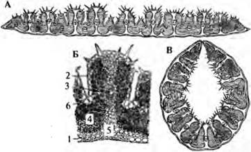
Leaves exhibit morphological and anatomical plasticity related to their ADAPTATION TO ENVIRONMENTAL conditions. For instance, in mesomorphic plants adapted to moderately moist (mesophytic) habitats, as well as in hydromorphic or aquatic plants, the walls of epidermal cells are thin. In contrast, xeromorphic plants thriving in arid (xerophytic) conditions feature an epidermis with thick, lignified walls covered by a robust cuticle, with stomata located in crypts sheltered by trichomes (Fig. 1.44, E). The epidermis in certain plants of arid regions is multi-layered, incorporating internal water-storage layers (Fig. 1.44, D, E). In some steppe grasses (Fig. 1.46), the epidermis contains motor or bulliform cells capable of rapidly releasing water, which causes the leaf to roll into a tube. This positions the stomata on the inside, thereby reducing evaporation.
Fig. 1.46. Feather grass leaf in cross section: A, B — expanded (under adequate moisture conditions); C — rolled (during moisture deficit); 1 — lower epidermis without stomata; 2 — upper, corrugated side, epidermis with stomata and trichomes; 3 — veins; 4 — chlorenchyma; 5 — sclerenchyma; 6 — motor cells
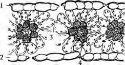
In hydrophytes and hygrophytes (Fig. 1.47), the mesophyll consists of aerenchyma, and stomata are found exclusively in the upper epidermis (epistomatic leaf). Shaded leaves often lack palisade parenchyma, or it is poorly developed, or consists of "umbent" parenchyma (where cells lie parallel to the leaf surface and transverse to the leaf blade, as in lily of the valley). In tropical grasses, bundle sheath cells are large (Fig. 1.48) and oriented such that their longitudinal axes are perpendicular to the bundle.
Fig. 1.47. Fragment of a water lily leaf cross section: 1 — upper epidermis with thick cuticle and stomata; 2 — substomatal air chambers; 3 — multi-layered palisade mesophyll; 4 — branched sclereids; 5 — aerenchyma; 6 — lower epidermis with suberized cells
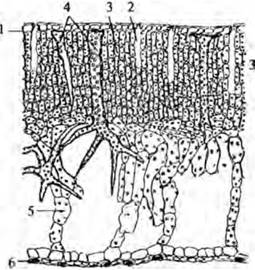
Fig. 1.48. Cytology/practical/72.html">Cross section of a barnyard grass leaf: 1, 2 — upper and lower epidermis; 3 — chlorenchyma; 4 — vein bundle sheath cells
Despite the plasticity of leaf structure, certain plant groups and species possess A number of fixed diagnostic features used in microscopic identification and analysis. These features include the type of stomatal apparatus, the type and Morphology of trichomes, their distribution pattern, and the presence, nature, and localization of crystalline inclusions, pigmented idioblasts, and secretory structures, among others.
Last update: 07/08/2026
Editorial and Educational Adaptation: This material has been compiled based on the primary/original source text. The project team performed an editorial review, corrected technical inaccuracies, structured sections, and adapted the content for an educational format.
What was processed:
- elimination of formatting defects (OCR errors, structural breaks, corrupted characters);
- editorial organization of content;
- standardization of terminology in accordance with academic sources;
- verification of factual statements against the original source text.
All mentions of the author, publication year, and origin of the primary text have been preserved in accordance with the source.