BIOLOGY Volume 1 - A Guide to General Biology - 2004
8. HETEROTROPHIC NUTRITION
8.3. The Human Alimentary Canal
8.3.2. The Human Dentition
Types of Teeth
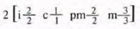
Humans have two jaws: a fixed upper jaw and a movable lower jaw. Both jaws are equipped with teeth, which are used to break down (chew) food. This mechanical process increases the surface area of the food exposed to the action of Enzymes. Teeth are extremely hard structures ideally adapted to perform their function. Humans develop two successive sets of teeth during their lifetime. The first to appear are the deciduous (milk) teeth, which are gradually replaced by the permanent teeth. Human teeth vary in shape, size, and the Morphology of their chewing surfaces. The total number of permanent teeth in an adult human is 32, comprising 8 incisors (i), 4 canines (c), 8 premolars (pm), and 12 molars (m). The dental arrangement is conventionally expressed by a dental formula. For permanent human teeth, it takes the following form:
Class="center">
where the letters represent the initial letters of the Latin names of the teeth; the number above the line indicates the number of teeth of each type in the upper jaw, and the number below the line indicates those in the lower jaw on each side (Fig. 8.13).

Fig. 8.13. A. X-ray of a human HEAD showing a row of permanent teeth on one side. B. Frontal X-ray of a human head showing the complete set of permanent teeth. Dental formula:
The number, size, and shape of teeth are adapted to the type of diet consumed. The Structure AND Functions of each tooth type are described below:
1. Incisors, located at the front of the Oral Cavity, have flat, sharp edges used for biting off and "cutting" food (Fig. 8.13).
2. Canines are cone-shaped. In humans, they are relatively poorly developed, but they are very prominent in carnivores, where they serve to seize and kill prey, and subsequently tear it into pieces.
3. Premolars (bicuspids) have one or two roots and two cusps on the chewing surface. They are designed for crushing and grinding food, although in humans they may also be used for tearing.
4. Molars have multiple roots (three in the upper jaw and two in the lower). The chewing surface of a molar features four or five cusps. These teeth serve for crushing and grinding food. They are absent in the deciduous set of human teeth.
General plan of tooth structure
The visible part of the tooth, called the crown, is covered with enamel (Fig. 8.14)—the hardest substance in the body, which is relatively resistant to decay. The neck of the tooth is embedded in the gum (gingiva), and the ROOT is anchored in the jawbone. Beneath the enamel lies dentin, which forms the bulk of the tooth. Dentin is quite durable, although it is inferior to enamel in hardness and resistance to decay. It is permeated by numerous microscopic tubules containing cytoplasmic processes of odontoblasts—the Cells responsible for forming dentin. The pulp cavity of the tooth contains odontoblasts, sensory nerve endings, and Blood Vessels that supply nutrients to the living Tissues of the tooth and remove their Metabolic waste products.

Fig. 8.14. Vertical section of a premolar.
The root of the tooth is covered with cementum, a bone-like tissue. Numerous fibres (periodontal ligament), attached at one end to the cementum and at the other to the bone, securely hold the tooth in place. However, the tooth retains a slight degree of mobility, which reduces the likelihood of it fracturing during chewing.
Dental diseases
There are two main dental diseases: periodontal disease and dental caries. Both are caused by The formation of dental plaque, which is a mixture of Bacteria and salivary components. If allowed to accumulate, bacteria cause inflammation of the Gums—periodontal disease (periodontitis). Through interaction with certain Cell/6.html">Chemical Components of saliva, dental plaque becomes hard and calcified, forming calculus (tartar) deposits that cannot be removed by simple brushing. Certain bacteria living in plaque are capable of converting dietary sugars into acid, which causes dental caries (tooth decay) (Fig. 8.15).

Fig. 8.15. Development of dental plaque: A. Spherical bacteria (cocci) are the first to colonize the tooth and, by multiplying, form a film. B. Microorganisms invade this matrix, which consists of bacterial secretions and salivary substances. C. The microbial community grows and becomes increasingly complex; populations of rod-shaped and filamentous bacteria appear. D. In the "climax" community, highly unusual associations between various populations can be observed, including structures resembling corn cobs.
PERIODONTITIS. Periodontitis is a gum disease caused by microorganisms present in the Mouth as part of dental plaque, particularly in the area between the gum and the tooth. Neglecting oral hygiene creates favorable conditions for The Development of this disease. Initially, gum inflammation occurs. This inflammation is usually painless; however, if left untreated, it paves the way for the disease to progress further, potentially spreading to the tooth root and destroying the fibers that anchor the tooth in place. Eventually, the tooth becomes loose and may fall out.
DENTAL CARIES. The microorganisms found in dental plaque are capable of converting sugar into acid. At first, the enamel is slowly and painlessly broken down by this acid. However, once the dentin and pulp are affected, severe pain arises, and the condition poses a threat of tooth loss. Several factors play a crucial role in the development of dental caries. These include the prolonged consumption of sugar-rich foods, changes in saliva composition, poor oral hygiene, and low fluoride levels in drinking Water. The Prevention of Dental caries is aided by the fluoridation of drinking water and certain foods, such as milk; The Use of fluoride supplements for children; the use of fluoride toothpastes; meticulous oral hygiene; regular visits to a dentist and dental hygienist; and maintaining a balanced, healthy diet.
Last update: 06/08/2026
Editorial and Educational Adaptation: This material has been compiled based on the primary/original source text. The project team performed an editorial review, corrected technical inaccuracies, structured sections, and adapted the content for an educational format.
What was processed:
- elimination of formatting defects (OCR errors, structural breaks, corrupted characters);
- editorial organization of content;
- standardization of terminology in accordance with academic sources;
- verification of factual statements against the original source text.
All mentions of the author, publication year, and origin of the primary text have been preserved in accordance with the source.