BIOCHEMISTRY - L. Stryer - 1984
VOLUME 2
PART III. BIOSYNTHESIS OF MACROMOLECULAR PRECURSORS
CHAPTER 22. BIOSYNTHESIS OF NUCLEOTIDES
22.15. Deoxyribonucleotides Are Synthesized by Reduction of Ribonucleoside Diphosphates
We now turn to the synthesis of deoxyribonucleotides. These DNA precursors are formed by the reduction of ribonucleotides. The 2'-hydroxyl group of the ribose residue is replaced by a hydrogen atom.
In E. coli and mammalian Cells, the substrates for this reaction are ribonucleoside diphosphates. The overall stoichiometry of this reaction is as follows:
Ribonucleoside diphosphate + NADPH + H+ → Deoxyribonucleoside diphosphate + NADP++ H2O.
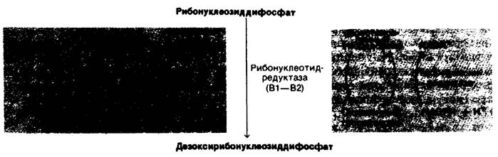
In reality, the reaction mechanism is much more complex than this equation suggests. Peter Reichard showed that in E. coli, electrons are transferred to the substrate through a series of sulfhydryl groups. The final reaction is catalyzed by Ribonucleotide reductase (also called ribonucleoside diphosphate reductase). The stoichiometry of this reaction is described by the following equation:
Class="center">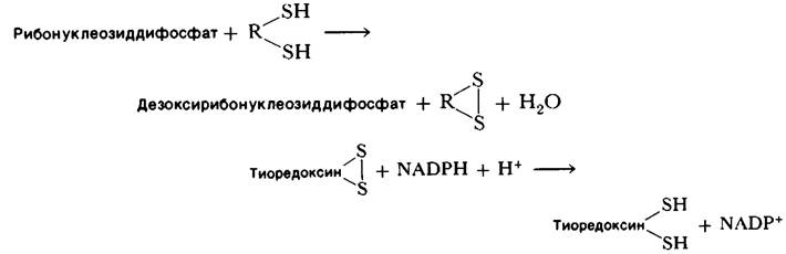
Fig. 22.17. Electron micrograph of aspartate transcarbamoylase
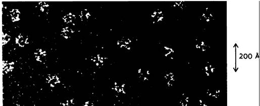
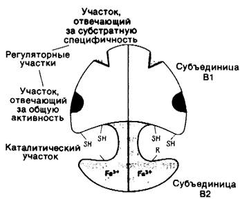
This enzyme consists of two subunits: B1 (a dimer with a molecular mass of 160 kDa) and B2 (a dimer with a molecular mass of 78 kDa). The B1 subunit contains binding sites for ribonucleotide substrates and allosteric effectors. In addition, B1 contains sulfhydryl groups that serve as immediate electron Donors in the reduction of the ribose residue. B2 is an iron-containing protein; it participates in catalysis, forming an unusual free radical on the aromatic ring of a Tyrosine residue. Subunits B1 and B2 together participate in The formation of the active sites of the enzyme (Fig. 22.18).
Fig. 22.18. Model of E. coli ribonucleotide reductase
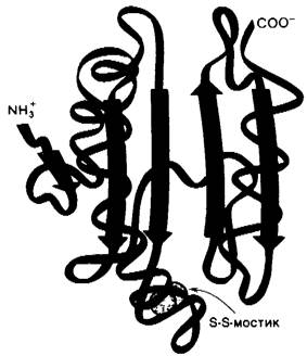
How are electrons transferred from NADPH to The sulfhydryl groups in the Active Site of ribonucleotide reductase? One of the Carriers of Reducing power is thioredoxin, a protein with a molecular mass of 12,000 Da containing two Cysteine residues in close proximity to each other (Fig. 22.19). These sulfhydryl groups are oxidized to form a disulfide in a reaction catalyzed by ribonucleotide reductase. Reduced thioredoxin is, in turn, regenerated by the reaction of NADPH with oxidized thioredoxin.
Fig. 22.19. Schematic representation of the conformation of the polypeptide backbone of oxidized E. coli thioredoxin. The reactive disulfide bond involved in the reaction is shown in yellow
This reaction is catalyzed by the flavoprotein thioredoxin reductase. It was formerly believed that thioredoxin was the sole carrier of reducing power to ribonucleotide reductase. However, an E. coli mutant completely lacking thioredoxin was found to synthesize deoxyribonucleotides. This unexpected finding led to the ISOLATION OF A second carrier system. The electron donor in this mutant proved to be Glutathione, a cysteine-containing tripeptide. As discussed above (Section 15.12), glutathione reductase catalyzes the reduction of oxidized glutathione (the disulfide form) by NADPH. In addition, another protein, glutaredoxin, is required to transfer the reducing power of glutathione to ribonucleotide reductase (Fig. 22.20). The relative contributions of the thioredoxin and glutaredoxin systems to ribonucleotide reduction in normal cells are not yet known.
Fig. 22.20. Ribonucleoside diphosphates are reduced to deoxyribonucleoside diphosphates by ribonucleotide reductase. Electrons are transferred from NADPH through a series of sulfhydryl groups. The sources of reducing power can be the thioredoxin system (shown in yellow) and the glutaredoxin system (shown in green)
The reduction of ribonucleoside diphosphates is precisely regulated by allosteric interactions. The B1 subunit of ribonucleotide reductase contains Two Types of allosteric sites: one regulates the overall activity of the enzyme, and the other regulates substrate Specificity. The overall catalytic activity of ribonucleotide reductase is decreased by the binding of dATP, which signals an Abundance of deoxyribonucleotides. This feedback inhibition is reversed by the binding of ATP. The binding of dATP or ATP to the substrate-specificity sites increases the reduction of the pyrimidine NUCLEOTIDES UDP and CDP. The reduction of GDP is stimulated by the binding of dTTP, which also inhibits the reduction of pyrimidine nucleotides. A subsequent increase in dGTP concentration leads to The stimulation of ADP reduction. Clearly, ribonucleotide reductase has multiple conformational states with different catalytic properties. This complex regulatory scheme ensures the synthesis of balanced amounts of the four deoxyribonucleotides for DNA Synthesis.
22.16. Deoxythymidylate Is Formed by Methylation of Deoxyuridylate
Uracil is not a component of DNA. Instead, DNA contains thymine, a methylated analog of uracil. The final Touch is added to The Structure of this precursor at the deoxyribonucleoside monophosphate level: deoxyuridylate (dUMP) is methylated to form deoxythymidylate (dTMP) by thymidylate synthase. The donor of the methyl group in this reaction is a tetrahydrofolate derivative, not S-adenosylmethionine. The methyl carbon comes from N5, N10-methylenetetrahydrofolate. Note that the methyl group introduced into deoxyuridylate is in a more reduced state than the methylene group in this tetrahydrofolate derivative. Where do the electrons for this reduction reaction come from? Two electrons are transferred from the tetrahydrofolate ring itself as a hydride ion (H-) that is cleaved from the ring. This hydrogen becomes part of the methyl group of dTMP. Tetrahydrofolate is oxidized to dihydrofolate in this reaction. Thus, N5, N10-methylenetetrahydrofolate serves as both the electron donor and the one-carbon unit donor in this methylation reaction (Fig. 22.21).
Fig. 22.21. Synthesis of dTMP from dUMP
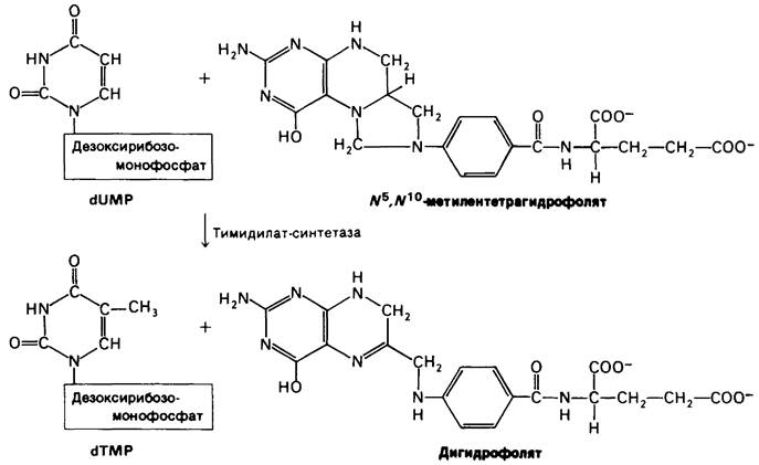
Recall that The transfer of a one-carbon unit occurs at the level of tetrahydrofolate, not dihydrofolate. Consequently, tetrahydrofolate must be regenerated. This is catalyzed by Dihydrofolate Reductase, using NADPH as a reducing agent:
Dihydrofolate + NADPH + H+ → Tetrahydrofolate + NADP+.
22.17. Some Anticancer Drugs Block the Synthesis of Deoxythymidylate
Rapidly dividing cells require large amounts of deoxythymidylate for DNA synthesis. The sensitivity of these cells to the inhibition of dTMP synthesis is exploited in Cancer Chemotherapy (Fig. 22.22). Typically, the Enzymes thymidylate synthase and dihydrofolate reductase are targeted. Fluorouracil (or fluorodeoxyuridine) is a widely used anticancer drug in clinical practice. It is converted in vivo to fluorodeoxyuridylate (F-dUMP). This dUMP analogue irreversibly inhibits thymidylate synthase after undergoing part of the catalytic cycle as a normal substrate. First, a sulfhydryl group of the enzyme adds to C-6 of the bound F-dUMP. Then, methylenetetrahydrofolate adds to C-5 of this intermediate. If dUMP participates in the reaction, the next step involves the transfer of a hydride ion to the methylene group of methylenetetrahydrofolate, and a proton is abstracted from C-5 of the bound nucleotide. However, if F-dUMP enters the reaction, the enzyme cannot Abstract F+, and catalysis is blocked at the stage of the covalent complex formation between F-dUMP, methylenetetrahydrofolate, and the sulfhydryl group of the enzyme.

Fig. 22.22. Thymidylate synthase and dihydrofolate reductase are targets of various drugs in cancer chemotherapy. Fluorodeoxyuridylate inhibits the methylation of dUMP. The Folic acid analogues aminopterin and amethopterin (methotrexate) block the regeneration of tetrahydrofolate.
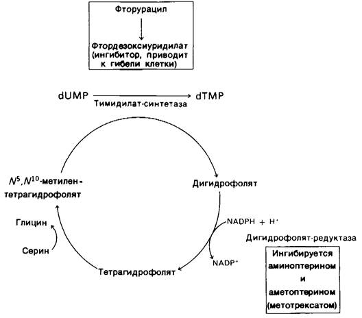
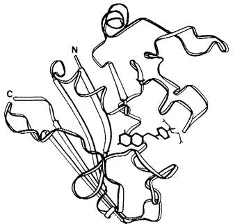
The synthesis of dTMP can also be blocked by inhibiting the regeneration of tetrahydrofolate (Fig. 22.22). Tetrahydrofolate analogues, such as aminopterin and amethopterin (methotrexate), are potent Competitive Inhibitors of dihydrofolate reductase (Ki < 10-9 M) (Fig. 22.24). Amethopterin is an important drug in the Treatment of acute leukemia and Choriocarcinoma.
Fig. 22.24. Three-dimensional structure of E. coli dihydrofolate reductase with a bound molecule of amethopterin (methotrexate)
Last update: 06/08/2026
Editorial and Educational Adaptation: This material has been compiled based on the primary/original source text. The project team performed an editorial review, corrected technical inaccuracies, structured sections, and adapted the content for an educational format.
What was processed:
- elimination of formatting defects (OCR errors, structural breaks, corrupted characters);
- editorial organization of content;
- standardization of terminology in accordance with academic sources;
- verification of factual statements against the original source text.
All mentions of the author, publication year, and origin of the primary text have been preserved in accordance with the source.