Biochemistry - The Chemical Reactions of Living Cells, Volume 3 - D. Metzler 1980
Biochemical Genetics and the Synthesis of Nucleic Acids and Proteins
Genetic Methods
Temperate Bacteriophages. Phage λ
When bacteriophage DNA penetrates a bacterial Cell, it almost instantaneously takes over The Cell's metabolic machinery and redirects it entirely toward producing new Viral Particles. As a result, within roughly 20 min, 100 to 200 new viral particles are formed, leading to cell lysis and death. Temperate phages behave in a fundamentally different manner. Upon entering the cell, the DNA of a temperate phage can be repressed and integrated with the bacterial genome much like the F factor (Fig. 15-2). It thus transitions into a prophage state and enters the so-called lysogenic phase of development: the repressed phage DNA replicates as part of the bacterial genome, doing no harm to the cell until some factor lifts the repression and "activates" the integrated genetic material. Following this, phage Replication and bacterial lysis take place. Temperate phages can also exist as Plasmids (e.g., phage P1).
The best-characterized temperate phage is phage lambda, which infects E. coli Cells [156—158]. Structurally, phage λ—which is characterized by a tail—somewhat resembles the T-even phages (Supplementary Material 4-D), yet its DNA genome is smaller: the Molecular Weight of phage λ DNA is approximately 31∙106, which corresponds to 46,500 Base Pairs. Within the bacterial cell, the ends of the phage λ DNA can join together to form a closed replicative form of the virus. In many infected cells (roughly 30%), phage λ DNA integrates into the bacterial chromosome at a specific site, att λ, located on the E. coli chromosome map at the position corresponding to 17 min. The integrated phage DNA constitutes a linear fragment representing about 1.2% of the total length of the E. coli chromosome. It replicates along with the rest of the chromosome and in most cases remains undetected.
Biochemists' interest in bacteriophage λ stems from several reasons. Most importantly, studying this bacteriophage offered hope of answering fundamental questions regarding the Regulation of METABOLISM/31.html">Transcription. We might ask, for instance, how it is that the majority of prophage λ genes can remain quiescent for many generations and then, under specific conditions, reignite the synthesis of active Viruses. The small size of the phage λ genome raised hopes that we could comprehend its Organization with considerable precision1). The host bacterium E. coli K12 is also exceptionally well-characterized genetically; furthermore, these Bacteria possess highly convenient amber suppressors that make it easy to identify bacteriophage Mutations. Moreover, the integrated prophage can undergo virtually any type of mutation, including large deletions, thereby enabling researchers to investigate complementation with other strains of this virus. A whole family of modified defective λ phages is known. Because the prophage occasionally captures neighboring GENES OF THE bacterial chromosome upon excision, researchers managed to isolate a group of transducing λ phages—that is, phages carrying specific genes of the bacterial chromosome and capable of transferring these genes to bacteria that lack them. The prospect of such Gene transfer into the genomes of plants and other higher organisms has sparked immense interest and heated debate. Another outcome of studying phage λ was The Development of a method allowing more precise mapping of gene positions.
a. Transcriptional Control
The answer to why phage λ genes can remain inactive for a given period came with the Discovery of the repressor protein [159, 161]. A single short Operon of prophage λ is constitutively transcribed by E. coli RNA polymerase. This operon contains the cI and rex genes. As shown in Fig. 15-22, transcription of these genes initiates from the l-strands of the prophage DNA. The protein encoded by the cI gene acts as a repressor. The repressor is an oligomer (most commonly a dimer) with a subunit molecular weight of 27,000. This protein binds to two operator Regions of the prophage DNA. One operator (oL) is located to the left and the other (oR) to the right of the cI gene (Fig. 15-22). Examination of DNA fragments protected by the repressor against nuclease attack revealed that each operator contains three subregions, which are sequentially filled by six repressor monomers (from left to right for oL and from right to left for oR). The nucleotide sequence of this region was deciphered, revealing that each of the hypothetical subregions possesses an axis of approximate twofold rotational Symmetry (i.e., it forms an almost palindromic sequence). Each subregion consists of 17 base pairs, with one half of the region containing the sequence TATCACCGC or a very similar one, whereas the other half is somewhat more variable. It is possible that when the dimeric repressor binds to each subregion, its monomers adopt quasi-equivalent Conformations [159].
1) The complete nucleotide sequence of phage λ has now been fully elucidated.—Ed. Note.
Class="center">
FIG. 15-22. Genetic and physical map of the phage λ genome [156]. A more detailed diagram of the Immunity region is given in Honigman A., Hu S-L., Chase R., Szybalski W., Nature (London), 262, 112—116 (1976).
By blocking these operators, the repressor prevents the synthesis of Enzymes required for the excision of phage λ DNA from the bacterial chromosome, as well as for the replication and Transcription of the remaining genes. The actual situation is more complex because establishing the initial lysogenic state apparently requires the products of the early left (cIII) and early right (cII) operon genes, which stimulate cI gene transcription. Once triggered, these genes function no further since they are never transcribed.
It is estimated that there are only a few molecules of phage λ repressor per cell. Under normal conditions, this is sufficient to maintain the prophage state. However, ultraviolet irradiation of the bacterium (acting presumably indirectly via the inhibition of DNA Synthesis) leads to the inactivation of the repressor and the transcription of other phage λ operons.
Transcription of the left operon begins at the pL promoter. The product of the first gene, N, is a protein that allows transcription to continue past the tL and tB terminators [161]. This protein is unstable, with a molecular half-life (t1/2) of approximately 2 min [162]. Leftward transcription proceeds through the exo and ß genes, which are involved in recombination, and the xis gene, which is required for the excision of the phage DNA from the chromosome. Upon Integration of the phage λ DNA, the E. coli chromosome breaks at the aa' sites (Fig. 15-22), and the DNA inserts immediately to the right of the gal operon (Fig. 15-1). Prophage transcription can now continue from the a' point into the bacterial genes. Introduction/27.html">Translation of the mRNA transcribed from this early left operon yields the enzymes necessary for prophage release and The formation of the circular replicative form of the phage DNA. DNA excision from the bacterial chromosome also occurs near the a' site; notably, during excision, phage λ can occasionally capture neighboring gal genes of the host cell.
The N gene product also enables rightward transcription through the O, P, and Q genes and subsequently along the rest of the chromosome to point a at a reduced rate. The O and P genes dictate the synthesis of Proteins that enable the host bacterium's replication machinery to initiate The production of new phage DNA molecules. Replication starts at the ori site and proceeds bidirectionally, as described in Section D. The Q gene directs the synthesis of a protein that drastically accelerates the transcription of late genes starting from the PR promoter.
As illustrated in Fig. 15-22, the chromosome is typically divided into four operons: a short repressor-producing operon, an early left operon, an early right operon, and a late operon1). The early operons primarily govern the synthesis of enzymes responsible for replication and recombination, as well as regulatory proteins. The late operon is associated with the synthesis of proteins required to assemble viral particles; it must be transcribed at a higher rate, which is ensured by the Q gene product. Within the late operon, genes A through F participate in phage λ DNA packaging and HEAD formation, whereas genes z through j govern tail synthesis and assembly. The S and R genes produce proteins that disrupt the host Cell Membrane and cause cell lysis. During the final Stages of the lytic developmental phase, most of the early genes are switched off by another phage λ repressor (encoded by the cro gene). As evident from the above, Transcriptional Regulation even in viruses can be a remarkably complex process.
1) A somewhat similar Gene Organization is found in T-even phages, phage T7, and other Bacteriophages.
b. "Sticky Ends" of DNA Molecules
While the replicative form of phage λ DNA is circular, the DNA of mature particles is famously linear. Unlike the linear DNA of T-even phages, phage λ DNA spontaneously forms either circles or linear "aggregates" upon release from the virion. This indicates that phage λ DNA possesses "sticky" ends that join together via specific base pairing. Direct nucleotide sequencing confirmed this assumption. Figure 15-23 illustrates the nucleotide sequence near the m and m' points (Fig. 15-22), where the replicative form of DNA unwinds, as well as at the sticky ends of the l- and r-strands. The circular form is thought to be opened by the action of an endonuclease. The existence of sticky ends is supported by more than just nucleotide sequencing data. It turned out that the two sites of hydrolytic bond Cleavage (marked by arrows in Fig. 15-23) are separated by 12 nucleotide pairs that form a DNA segment with a strikingly high degree of symmetry [163, 164]. Thus, we once again encounter a palindrome, which in this instance forms a specific recognition site within the DNA molecule.
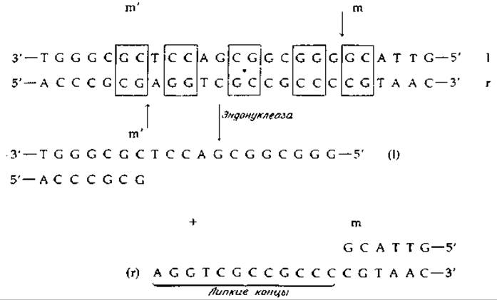
FIG. 15-23. Sticky ends of phage DNA and their generation from the replicative form by endonuclease action. Note the axis of local approximate twofold symmetry (its position marked by a bold dot), which serves as a specific interaction site for the symmetrical dimeric enzyme. Symmetrically positioned base pairs are boxed. The mm' points correspond to those on the genetic map (Fig. 15-22).
c. Heteroduplexes and Physical Gene Mapping
The availability of phages carrying significant deletions in various genome regions enabled the development of a novel gene-mapping method using direct Electron Microscopy observations [165]. First, DNA is isolated from two different phage strains, such as a wild-type strain X and a mutant strain with deletions in a specific gene or genes. The resulting DNA is readily denatured, after which the r- and l-strands can be separated. If the l-strands of one strain are mixed with the r-strands of another strain and subjected to annealing, double-stranded DNA formation will be observed. However, because one strain contains a deletion, the homologous region of normal phage λ DNA forms a single-stranded loop that can be easily visualized using an Electron microscope. Figure 15-24 shows an electron micrograph of such a heteroduplex molecule containing a deletion loop as an example. This photograph also reveals a "bubble" formed where a stretch of nonhomologous DNA was incorporated into one of the strands [165]. Because distances can be measured with high precision on electron micrographs, this approach allows for the construction of accurate "physical maps." The chromosomal map shown in Fig. 15-22 is such a "physical map," and therefore the distances indicated on it are more precise than those on the E. coli genetic map shown in Fig. 15-1. This technique has also been applied to study colicinogenic factors [166].

FIG. 15-24. A. Electron micrograph of a heteroduplex DNA molecule formed from complementary strands of λb2 and λimm 434 phages. In phage λb2, a segment of phage λ DNA forms a deletion loop (designated b2), whereas in phage λimm 434 a portion of the DNA is replaced by phage λ DNA, generating a "nonhomology bubble" (designated i 434/iλ). The sticky ends of the DNA are labeled v. e. B. Higher magnification of the nonhomology bubble. C. Schematic drawing explaining the image shown in panel B. The arrow points to a short (20–150 NUCLEOTIDES) homologous region (Westmoreland B. C. et al., Science, 163, 1343–1348, 1969).
Last update: 06/08/2026
Editorial and Educational Adaptation: This material has been compiled based on the primary/original source text. The project team performed an editorial review, corrected technical inaccuracies, structured sections, and adapted the content for an educational format.
What was processed:
- elimination of formatting defects (OCR errors, structural breaks, corrupted characters);
- editorial organization of content;
- standardization of terminology in accordance with academic sources;
- verification of factual statements against the original source text.
All mentions of the author, publication year, and origin of the primary text have been preserved in accordance with the source.